Skip to main content
Thank you for visiting nature.com. You are using a browser version with limited support for CSS. To obtain
the best experience, we recommend you use a more up to date browser (or turn off compatibility mode in
Internet Explorer). In the meantime, to ensure continued support, we are displaying the site without styles
and JavaScript.
Advertisement


●
●
●
●
●
●
●
●
●
(一)nature
(二)articles
(三)
article
●Article
●Published:
X-ray analysis on the nanogram to microgram scale using porous complexes
●Yasuhide Inokuma1,
●Shota Yoshioka1,
●Junko Ariyoshi1,
●Tatsuhiko Arai1,
●Yuki Hitora2,
●Kentaro Takada2,
●Shigeki Matsunaga2,
●Kari Rissanen3 &
●…
●Makoto Fujita1
Nature
volume 495, pages 461–466 (2013)Cite this article
93k Accesses
704 Citations
185 Altmetric
Metrics details
Subjects
●Biochemistry
●Coordination chemistry
●X-ray diffraction
ACorrigendum to this article was published on 21 August 2013
Abstract
X-ray single-crystal diffraction (SCD) analysis has the intrinsic limitation that the target molecules must be obtained as single crystals. Here we report a protocol for SCD analysis that does not require the crystallization of the sample. In our method, tiny crystals of porous complexes are soaked in a solution of the target, such that the complexes can absorb the target molecules. Crystallographic analysis clearly determines the absorbed guest structures along with the host frameworks. Because the SCD analysis is carried out on only one tiny crystal of the complex, the required sample mass is of the nanogram–microgram order. We demonstrate that as little as about 80 nanograms of a sample is enough for the SCD analysis. In combination with high-performance liquid chromatography, our protocol allows the direct characterization of multiple fractions, establishing a prototypical means of liquid chromatography SCD analysis. Furthermore, we unambiguously determined the structure of a scarce marine natural product using only 5 micrograms of the compound.
Access through your institution
Buy or subscribe
This is a preview of subscription content, access via your institution
Access options
Access through your institution
Access through your institution
Change institution
Buy or subscribe
Subscription info for Japanese customers
We have a dedicated website for our Japanese customers. Please go to natureasia.com to subscribe to this journal.
Go to natureasia.com
Buy this article
●Purchase on Springer Link
●Instant access to full article PDF
Buy now
Prices may be subject to local taxes which are calculated during checkout
Additional access options:
●
Log in
●
Learn about institutional subscriptions
●
Read our FAQs
●
Contact customer support
Figure 1: X-ray crystallographic observation of liquid guest molecules using crystalline sponges.
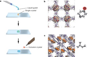 Figure 2: Nanogram-scale guest inclusion with a crystal of crystalline sponge3.
 Figure 3: Crystal structures of a variety of guests determined using a one-crystal-scale inclusion protocol.
 Figure 4: The crystal structure of a chiral guest, santonin, trapped in a crystalline sponge.
 Figure 5: LC–SCD analysis of natural flavonoids.
 Figure 6: Structural determination of miyakosyne A.

Article
Open access
30 January 2023
Article
Open access
02 October 2019
Article
27 July 2022
References
Ooi, L. Principles of X-Ray Crystallography (Oxford Univ. Press, 2010)
Google Scholar
Sheldrick, G. M. A short history of SHELX . Acta Crystallogr. A 64, 112–122 (2008)
Article
ADS
CAS
Google Scholar
Ohashi, Y. in Models, Mysteries and Magic of Molecules (eds Boeyens, J. C. A. & Ogilvie, J. F. ) 109–113 (Springer, 2008)
Book
Google Scholar
Batten, S. R. & Robson, R. Interpenetrating nets: ordered, periodic entanglement. Angew. Chem. Int. Ed. 37, 1460–1494 (1998)
Article
Google Scholar
Kitagawa, S., Kitaura, R. & Noro, S. Functional porous coordination polymers. Angew. Chem. Int. Ed. 43, 2334–2375 (2004)
Article
CAS
Google Scholar
Yaghi, O. M. et al. Reticular synthesis and the design of new materials. Nature 423, 705–714 (2003)
Article
ADS
CAS
Google Scholar
Fujita, M., Kwon, Y. J., Washizu, S. & Ogura, K. Preparation, clathration ability and catalysis of a two-dimensional square network material composed of cadmium(II) and 4,4′-bipyridine. J. Am. Chem. Soc. 116, 1151–1152 (1994)
Article
CAS
Google Scholar
Inokuma, Y., Arai, T. & Fujita, M. Networked molecular cages as crystalline sponges for fullerenes and other guests. Nature Chem. 2, 780–783 (2010)
Article
ADS
CAS
Google Scholar
Biradha, K. & Fujita, M. A springlike 3D-coordination network that shrinks or swells in a crystal-to-crystal manner upon guest removal or readsorption. Angew. Chem. Int. Ed. 41, 3392–3395 (2002)
Article
CAS
Google Scholar
Fujita, M. et al. Self-assembly of ten molecules into nanometre-sized organic host frameworks. Nature 378, 469–471 (1995)
Article
ADS
CAS
Google Scholar
Inokuma, Y., Kojima, N., Arai, T. & Fujita, M. Bimolecular reaction via the successive introduction of two substrates into the crystals of networked molecular cages. J. Am. Chem. Soc. 133, 19691–19693 (2011)
Article
CAS
Google Scholar
Ohmori, O., Kawano, M. & Fujita, M. Crystal-to-crystal guest exchange of large organic molecules within a 3D coordination network. J. Am. Chem. Soc. 126, 16292–16293 (2004)
Article
CAS
Google Scholar
Haneda, T., Kawano, M., Kojima, T. & Fujita, M. Thermo-to-photo-switching of the chromic behavior of salicylideneanilines by inclusion in a porous coordination network. Angew. Chem. Int. Ed. 46, 6643–6645 (2007)
Article
CAS
Google Scholar
Ohara, K., Kawano, M., Inokuma, Y. & Fujita, M. A porous coordination network catalyzes an olefin isomerization reaction in the pore. J. Am. Chem. Soc. 132, 30–31 (2010)
Article
CAS
Google Scholar
Férey, G. Hybrid porous solids: past, present, future. Chem. Soc. Rev. 37, 191–214 (2008)
Article
Google Scholar
Li, J.-R., Kuppler, R. J. & Zhou, H.-C. Selective gas adsorption and separation in metal–organic frameworks. Chem. Soc. Rev. 38, 1477–1504 (2009)
Article
CAS
Google Scholar
Chen, B., Xiang, S. & Qian, G. Metal-organic frameworks with functional pores for recognition of small molecules. Acc. Chem. Res. 43, 1115–1124 (2010)
Article
CAS
Google Scholar
Kondo, M. et al. Three-dimensional framework with channeling cavities for small molecules: {[M2(4,4′-bpy)3(NO3)4]•xH2O} n(M = Co, Ni, Zn). Angew. Chem. Int. Edn Engl. 36, 1725–1727 (1997)
Article
CAS
Google Scholar
Yoshizawa, M., Klosterman, J. K. & Fujita, M. Functional molecular flasks: new properties and reactions within discrete, self-assembled hosts. Angew. Chem. Int. Ed. 48, 3418–3438 (2009)
Article
CAS
Google Scholar
Inokuma, Y., Kawano, M. & Fujita, M. Crystalline molecular flasks. Nature Chem. 3, 349–358 (2011)
Article
ADS
CAS
Google Scholar
Li, Q. W. et al. Docking in metal-organic frameworks. Science 325, 855–859 (2009)
Article
ADS
CAS
Google Scholar
Kim, H., Chun, H., Kim, G.-H., Lee, H.-S. & Kim, K. Vapor phase inclusion of ferrocene and its derivative in a microporous metal-organic porous material and its structural characterization by single crystal X-ray diffraction. Chem. Commun. 2759–2761 (2006)
Halder, G. J. & Kepert, C. J. In situ single-crystal X-ray diffraction studies of desorption and sorption in a flexible nanoporous molecular framework material. J. Am. Chem. Soc. 127, 7891–7900 (2005)
Article
CAS
Google Scholar
Kawano, M. & Fujita, M. Direct observation of crystalline-state guest exchange in coordination networks. Coord. Chem. Rev. 251, 2592–2605 (2007)
Article
CAS
Google Scholar
Kitaura, R. et al. Formation of a one-dimensional array of oxygen in a microporous metal-organic solid. Science 298, 2358–2361 (2002)
Article
ADS
CAS
Google Scholar
Cahn, R. S., Ingold, C. & Prelog, V. Specification of molecular chirality. Angew. Chem. Int. Edn Engl. 5, 385–415 (1966)
Article
CAS
Google Scholar
Seco, J. M., Quiñoá, E. & Riguera, R. The assignment of absolute configuration by NMR. Chem. Rev. 104, 17–118 (2004)
Article
CAS
Google Scholar
Freedman, T. B., Cao, X., Dukor, R. K. & Nafie, L. A. Absolute configuration determination of chiral molecules in the solution state using vibrational circular dichroism. Chirality 15, 743–758 (2003)
Article
CAS
Google Scholar
Bijvoet, J. M., Peerdeman, A. F. & van Bommel, A. J. Determination of the absolute configuration of optically active compounds by means of X-rays. Nature 168, 271–272 (1951)
Article
ADS
CAS
Google Scholar
Flack, H. D. & Bernardinelli, G. Absolute structure and absolute configuration. Acta Crystallogr. A 55, 908–915 (1999)
Article
CAS
Google Scholar
Corey, E. J. The stereochemistry of santonin, β-santonin, and artemisin. J. Am. Chem. Soc. 77, 1044–1045 (1955)
Article
CAS
Google Scholar
Deschamps, J. R. X-ray crystallography of chemical compounds. Life Sci. 86, 585–589 (2010)
Article
CAS
Google Scholar
Takayanagi, H., Sudou, M. & Ogura, H. Crystal structure of 1α,2β-dibromo-1,2-dihydro-α-santonin. Anal. Sci. 7, 183–184 (1991)
Article
CAS
Google Scholar
Inayama, S. et al. Unusual bromination of tetrahydro-(–)-α-santonins and new santonin isomers: X-ray crystal and molecular structure of 2β,14-dibromo-4α,5β,6β,11βH-tetrahydrosantonin. J. Chem. Soc. Chem. Commun. 495–496. (1980)
Green, C. O., Wheatley, A. O., Osagie, A. U., Morrison, E. Y. S. A. & Asemota, H. N. Determination of polymethoxylated flavones in peels of selected Jamaican and Mexican citrus (Citrus spp.) cultivars by high-performance liquid chromatography. Biomed. Chromatogr. 21, 48–54 (2007)
Article
CAS
Google Scholar
Han, S. et al. Isolation and identification of polymethoxyflavones from the hybrid Citrus, Hallabong. J. Agric. Food Chem. 58, 9488–9491 (2010)
Article
CAS
Google Scholar
Hitora, Y., Takada, K., Okada, S. & Matsunaga, S. Miyakosynes A–F, cytotoxic methyl branched acetylenes from a marine sponge Petrosia sp. Tetrahedron 67, 4530–4534 (2011)
Article
CAS
Google Scholar
Sampietro, D. A., Catalan, C. A. N. & Vattuone, M. A. Isolation, Identification, and Characterization of Allelochemicals/Natural Products (Science Publ., 2009)
Book
Google Scholar
Croue, J.-P., Korshin, G. V. & Benjamin, M. M. Characterization of Natural Organic Matter in Drinking Water 73–374 (Am. Water Works Assoc., 1999)
Google Scholar
Ahuja, S. & Alsante, K. Handbook of Isolation and Characterization of Impurities in Pharmaceuticals (Academic, 2003)
Google Scholar
Download references
Acknowledgements
This research was supported by Grants-in-Aid for Specially Promoted Research (24000009) and Young Scientists (B) (23750146), and by the CREST project of the Japan Science and Technology Agency. The experiment involving X-ray crystallography with 80 ng of guest molecules was performed using VariMax optics with a RAPID image plate detector system, courtesy of Rigaku Corporation. We thank M. Yamasaki and H. Sato for support for X-ray measurements.
Author information
Authors and Affiliations
Department of Applied Chemistry, Graduate School of Engineering, The University of Tokyo, Hongo, Bunkyo-ku, Tokyo, 113-8656, Japan,
Yasuhide Inokuma, Shota Yoshioka, Junko Ariyoshi, Tatsuhiko Arai & Makoto Fujita
Laboratory of Aquatic Natural Products Chemistry, Graduate School of Agricultural and Life Science, The University of Tokyo, Yayoi, Bunkyo-ku, Tokyo, 113-8657, Japan ,
Yuki Hitora, Kentaro Takada & Shigeki Matsunaga
Department of Chemistry, NanoScience Center, University of Jyväskylä, PO Box 35, 40014 Jyväskylä, Finland,
Kari Rissanen
Authors
(一)Yasuhide Inokuma
View author publications
You can also search for this author in
PubMed Google Scholar
(二)Shota Yoshioka
View author publications
You can also search for this author in
PubMed Google Scholar
(三)Junko Ariyoshi
View author publications
You can also search for this author in
PubMed Google Scholar
(四)Tatsuhiko Arai
View author publications
You can also search for this author in
PubMed Google Scholar
(五)Yuki Hitora
View author publications
You can also search for this author in
PubMed Google Scholar
(六)Kentaro Takada
View author publications
You can also search for this author in
PubMed Google Scholar
(七)Shigeki Matsunaga
View author publications
You can also search for this author in
PubMed Google Scholar
(八)Kari Rissanen
View author publications
You can also search for this author in
PubMed Google Scholar
(九)Makoto Fujita
View author publications
You can also search for this author in
PubMed Google Scholar
Contributions
Y.I. and M.F. designed the project, analysed results and wrote the manuscript. S.Y., J.A. and T.A. performed the experimental work and crystallographic analysis. Y.H., S.M. and K.T. selected and provided a natural product sample for analysis. K.R. confirmed the validity of the X-ray crystallographic analysis of all data.
Corresponding author
Correspondence to
Makoto Fujita.
Ethics declarations
Competing interests
The authors declare no competing financial interests.
Additional information
The X-ray crystallographic coordinates for structures reported in this paper have been deposited at the Cambridge Crystallographic Data Centre, under deposition numbers CCDC 910380, 910381, 910382, 910383, 910384, 910385, 910386, 910387, 910388, 910389, 910390, 910391, 910392, 910393 and 910394. These data can be obtained free of charge from the Cambridge Crystallographic Data Centre (http://www.ccdc.cam.ac.uk/data_request/cif).
Supplementary information
This file contains Supplementary Methods, Supplementary Text and Data Supplementary Figures 1-5 and additional references. (PDF 3503 kb)
This file contains the crystallographic data. This file was added online on 8 April, 2013. (TXT 774 kb)
PowerPoint slides
Rights and permissions
Reprints and permissions
About this article
Cite this article
Inokuma, Y., Yoshioka, S., Ariyoshi, J. et al. X-ray analysis on the nanogram to microgram scale using porous complexes.
Nature 495, 461–466 (2013). https://doi.org/10.1038/nature11990
Download citation
Received:
Accepted:
Published:
Issue Date:
DOI: https://doi.org/10.1038/nature11990
Share this article
Anyone you share the following link with will be able to read this content:
Sorry, a shareable link is not currently available for this article.
Provided by the Springer Nature SharedIt content-sharing initiative
This article is cited by
●Yuki Wada
●Pavel M. Usov
●Masaki Kawano
Nature Communications (2024)
●Daiji Ogata
●Shota Koide
●Junpei Yuasa
Nature Communications (2024)
●Emily C. Gentry
●Stephanie L. Collins
●Pieter C. Dorrestein
Nature (2024)
●Gabin Thierry M. Bitchagno
●Vaderament-A. Nchiozem-Ngnitedem
●Serge Alain Fobofou
Nature Reviews Chemistry (2022)
●Tomasz Poręba
●Piero Macchi
●Michelle Ernst
Nature Communications (2022)
Comments
By submitting a comment you agree to abide by our Terms and Community Guidelines. If you find something abusive or that does not comply with our terms or guidelines please flag it as inappropriate.
Access through your institution
Buy or subscribe
Access through your institution
Change institution
Buy or subscribe
Editorial Summary
Crystal structure without the crystals
X-ray single crystal diffraction provides direct structural information of molecules at the atomic level and is recognized as a reliable structure determination method. However, as its name implies, the technique has a limitation, the sample needs to be available as a single crystal, the growth of which can be a time consuming process of trial-and-error. This paper describes a new X-ray analysis protocol that does not require crystallization of the sample itself. Instead, crystalline 'sponges' known as metal organic frameworks are used to soak up a drop of a liquid guest containing the target molecule. The sponges contain pores that recognize the target molecules and bind them in an ordered array, enabling the crystallographic structure analysis of the absorbed guest along with the host framework. The method is demonstrated with the absolute structure determination of a scarce natural product, miyakosyne A, using little more than a trace, 5 μg of sample. Corrected 8 April 2013
Associated content
●Pierre Stallforth
●Jon Clardy
Nature
News & Views
●Yasuhide Inokuma
●Shota Yoshioka
●Makoto Fujita
Nature Protocols
Protocol
2014
Advertisement

●
●
●
●
●
●
●
●
●
●
●
●
●
●
●
●
●
●
●
●
●
●
●
●
●
●
●
●
●
●
●
●
●
●
●
●
Nature (Nature)
ISSN 1476-4687 (online)
ISSN 0028-0836 (print)
nature.com sitemap
●
●
●
●
●
●
●
●
●
●
●
●
●
●
●
●
●
●
●
●
●
●
●
●
●
●
●
●
●
●
●
●
●
●
●
kies
●
●
●
●
© 2024 Springer Nature Limited

Sign up for the Nature Briefing newsletter — what matters in science, free to your inbox daily.
Get the most important science stories of the day, free in your inbox.
Sign up for Nature Briefing


