Jump to content


Main menu


Navigation  


Main page
Contents
Current events
Random article
About Wikipedia
Contact us
Donate


Contribute  


Help
Learn to edit
Community portal
Recent changes
Upload file


Search  


Create account

Log in


Create account
 Log in


Pages for logged out editors learn more  


Contributions
Talk


Contents


(Top)


1 Structure  


1.1  Development  


2 Function  


3 Clinical significance  


4 Disease  


5 See also  


6 Additional images  


7 Notes  


8 References  


9 External links  


Calcaneus


العربية
Azərbaycanca
Башҡортса
Bosanski
Brezhoneg
Català
Чӑвашла
Čeština
Deutsch
ދިވެހިބަސް
Eesti
Ελληνικά
Español
Esperanto
Euskara
فارسی
Français
Galego

Hrvatski
Ido
Italiano
Latina
Lingua Franca Nova
Lombard
Nederlands

Polski
Português
Română
Русский
Sicilianu
Simple English
Slovenčina
Srpskohrvatski / српскохрватски
Suomi
Svenska
Türkçe
Українська
 

Edit links


Article
Talk


Read
Edit
View history


Tools


Actions  


Read
Edit
View history


General  


What links here
Related changes
Upload file
Special pages
Permanent link
Page information
Cite this page
Get shortened URL
Download QR code
Wikidata item


Print/export  


Download as PDF
Printable version


In other projects  


Wikimedia Commons


Appearance


From Wikipedia, the free encyclopedia
 

(Redirected from Calcaneum)

Calcaneus

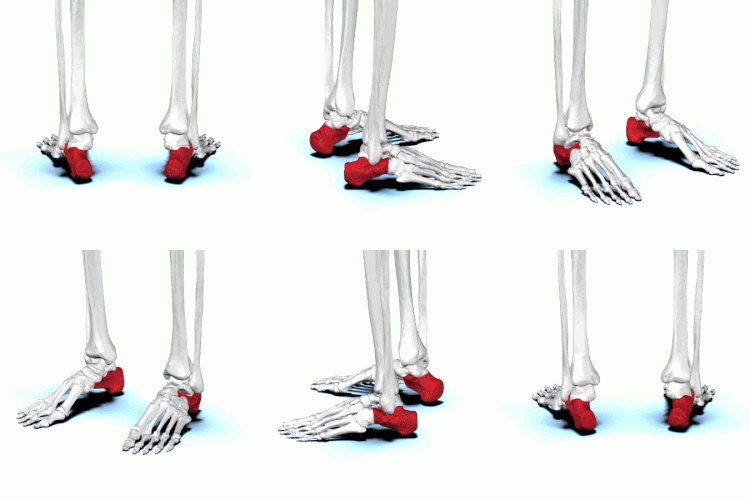
The calcaneus forms the bony part of the heel. It forms a joint with the talus bone, the subtalar joint.
Bones of the foot, with the calcaneus shown in red

Details

Identifiers

Latin

calcaneus, calcaneum, os calcis

MeSH

D002111

TA98

A02.5.11.001

TA2

1468

FMA

24496

Anatomical terms of bone

[edit on Wikidata]

In humans and many other primates, the calcaneus (/kælˈkniəs/; from the Latin calcaneusorcalcaneum, meaning heel;[1] pl.: calcaneiorcalcanea) or heel bone is a bone of the tarsus of the foot which constitutes the heel. In some other animals, it is the point of the hock.

Structure[edit]

In humans, the calcaneus is the largest of the tarsal bones and the largest bone of the foot. Its long axis is pointed forwards and laterally.[2] The talus bone, calcaneus, and navicular bone are considered the proximal row of tarsal bones.[3] In the calcaneus, several important structures can be distinguished:[3]

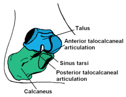
There is a large calcaneal tuberosity located posteriorly on plantar surface with medial and lateral tubercles on its surface. Besides, there is another peroneal tubercle on its lateral surface.[2] On its lower edge on either side are its lateral and medial processes (serving as the origins of the abductor hallucis and abductor digiti minimi). The Achilles tendon is inserted into a roughened area on its superior side and the cuboid bone articulates with its anterior side.[citation needed] On its superior side there are three articular surfaces for the articulation with the talus bone.[2] Between these superior articulations and the equivalents on the talus is the tarsal sinus (a canal occupied by the interosseous talocalcaneal ligament).[citation needed] At the upper and forepart of the medial surface of the calcaneus, below the middle talar facet, there is a horizontal eminence, the talar shelf (also sustentaculum tali).[2] Sustentaculum tali gives attachment to the plantar calcaneonavicular (spring) ligament, tibiocalcaneal ligament, and medial talocalcaneal ligament. This eminence is concave above, and articulates with the middle calcaneal articular surface of the talus; below, it is grooved for the tendon of the flexor hallucis longus; its anterior margin gives attachment to the plantar calcaneonavicular ligament, and its medial margin to a part of the deltoid ligament of the ankle-joint.

On the lateral side is commonly a tubercle called the calcaneal tubercle (or trochlear process). This is a raised projection located between the tendons of the peroneus longus and brevis. It separates the two oblique grooves of the lateral surface of the calcaneus (for the tendons of the peroneal muscles).

Its chief anatomical significance is as a point of divergence of the previously common pathway shared by the distal tendons of peroneus longus and peroneus brevis en route to their distinct respective attachment sites. [3]

The calcaneus is part of two joints: the proximal intertarsal joint and the talocalcaneal joint. The point of the calcaneus is covered by the calcanean bursa.

Development[edit]

In the calcaneus, an ossification center develops during the 4th7th weekoffetal development. [3]

Function[edit]

Three muscles insert on the calcaneus: the gastrocnemius, soleus, and plantaris. These muscles are part of the posterior compartment of the leg and aid in walking, running and jumping. Their specific functions include plantarflexion of the foot, flexion of the knee, and steadying the leg on the ankle during standing. The calcaneus also serves as origin for several short muscles that run along the sole of the foot and control the toes.

Muscle attachments (seen from above)

Muscle attachments (seen from below)

Muscle

Direction

Attachment[4]

Gastrocnemius

Insertion

Calcaneal tubercle through the achilles tendon

Soleus

Insertion

Calcaneal tubercle through the achilles tendon

Plantaris

Insertion

Calcaneal tubercle either directly or through the achilles tendon

Extensor digitorum brevis

Origin

Dorsal side of calcaneus

Abductor hallucis

Origin

Medial process of calcaneus

Extensor hallucis brevis

Origin

Dorsal side of calcaneus

Abductor digiti minimi

Origin

Calcaneal tubercle

Flexor digitorum brevis

Origin

Calcaneal tubercle

Quadratus plantae

Origin

Lateral and medial processes of calcaneus
Clinical significance[edit]

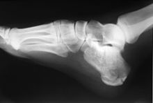
Calcaneus fracture X-ray

Normally the tibia sits vertically above the calcaneus (pes rectus). If the calcaneal axis between these two bones is turned medially the foot is in an everted position (pes valgus), and if it is turned laterally the foot is in an inverted position (pes varus).[5]

Disease[edit]

The talar shelf is typically involved in subtalar or talocalcaneal tarsal coalition.

See also[edit]

Additional images[edit]

Notes[edit]

  1. ^ Mosby’s Medical, Nursing and Allied Health Dictionary, Fourth Edition, Mosby-Year Book Inc., 1994, p. 242
  • ^ a b c d Ryan, Stephanie (2011). "Chapter 8". Anatomy for diagnostic imaging (Third ed.). Elsevier Ltd. p. 284. ISBN 9780702029714.
  • ^ a b c d Platzer (2004), p 216
  • ^ Bojsen-Møller, Finn; Simonsen, Erik B.; Tranum-Jensen, Jørgen (2001). Bevægeapparatets anatomi [Anatomy of the Locomotive Apparatus] (in Danish) (12th ed.). pp. 364–367. ISBN 978-87-628-0307-7.
  • ^ Thieme Atlas of Anatomy (2006), p 410
  • References[edit]

    External links[edit]

    Femur

    upper extremity

  • neck
  • greater trochanter
  • lesser trochanter
  • intertrochanteric line
  • intertrochanteric crest
  • quadrate tubercle
  • shaft

  • upper intermediate: pectineal line
  • upper lateral: gluteal tuberosity / third trochanter
  • lower extremity

  • patellar surface
  • epicondyles
  • condyles
  • intercondylar fossa
  • Tibia

    upper extremity

  • condyles
  • medial
  • intercondylar area
  • shaft

  • soleal line
  • lower extremity

  • Posterior colliculus
  • fibular notch
  • Fibula

    Other

    Foot

    Tarsus

  • calcaneal tubercle
  • talus
  • navicular
  • cuboid
  • cuneiform
  • Metatarsals

  • 2nd
  • 3rd
  • 4th
  • 5th
  • Other

    National

  • BnF data
  • Germany
  • Israel
  • United States
  • Czech Republic
  • Other


    Retrieved from "https://en.wikipedia.org/w/index.php?title=Calcaneus&oldid=1211079443"

    Categories: 
    Bones of the lower limb
    Vertebrate anatomy
    Bones of the foot
    Tarsal bones
    Hidden categories: 
    CS1 Danish-language sources (da)
    Articles with short description
    Short description is different from Wikidata
    All articles with unsourced statements
    Articles with unsourced statements from April 2022
    Commons category link is on Wikidata
    Articles with BNF identifiers
    Articles with BNFdata identifiers
    Articles with GND identifiers
    Articles with J9U identifiers
    Articles with LCCN identifiers
    Articles with NKC identifiers
    Articles with TA98 identifiers


    This page was last edited on 29 February 2024, at 19:11 (UTC).

    Text is available under the Creative Commons Attribution-ShareAlike License 4.0; additional terms may apply. By using this site, you agree to the Terms of Use and Privacy Policy. Wikipedia® is a registered trademark of the Wikimedia Foundation, Inc., a non-profit organization.


    Privacy policy

    About Wikipedia

    Disclaimers

    Contact Wikipedia

    Code of Conduct

    Developers

    Statistics

    Cookie statement

    Mobile view


    Wikimedia Foundation
    Powered by MediaWiki