Jump to content


Main menu


Navigation  


Main page
Contents
Current events
Random article
About Wikipedia
Contact us
Donate


Contribute  


Help
Learn to edit
Community portal
Recent changes
Upload file


Search  


Create account

Log in


Create account
 Log in


Pages for logged out editors learn more  


Contributions
Talk


Contents


(Top)


1 References  


Fascia dentata


Add links


Article
Talk


Read
Edit
View history


Tools


Actions  


Read
Edit
View history


General  


What links here
Related changes
Upload file
Special pages
Permanent link
Page information
Cite this page
Get shortened URL
Download QR code
Wikidata item


Print/export  


Download as PDF
Printable version


Appearance


From Wikipedia, the free encyclopedia


Fascia dentata
Coronal section of inferior horn of lateral ventricle. (Fascia dentata labeled at bottom left.)
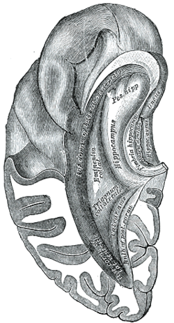
Inferior and posterior cornua, viewed from above. (Fascia dentata labeled at center right.)
Anatomical terms of neuroanatomy

[edit on Wikidata]

The fascia dentata is the earliest stage of the hippocampal circuit.[1][2] Its primary input is the perforant path from the superficial layers of entorhinal cortex. Its principal neurons are tiny granule cells which give rise to unmyelinated axons called the mossy fibers which project to the hilus and CA3. The fascia dentata of the rat contains approximately 1,000,000 granule cells. It receives feedback connections from mossy cells in the hilus at distant levels in the septal and temporal directions. The fascia dentata and the hilus together make up the dentate gyrus. As with all regions of the hippocampus, the dentate gyrus also receives GABAergic and cholinergic input from the medial septum and the diagonal band of Broca.

References

[edit]
  1. ^ Zimmer, Jens (1978-01-01), Corner, M. A.; Baker, R. E.; Vandepoll, N. E.; Swaab, D. F. (eds.), "Development of the Hippocampus and Fascia Dentata: Morphological and Histochemical Aspects", Progress in Brain Research, Maturation of the Nervous System, vol. 48, Elsevier, pp. 171–190, doi:10.1016/s0079-6123(08)61022-5, ISBN 978-0-444-80036-7, PMID 370906, retrieved 2023-03-29
  • ^ Zimmer, J.; Gähwiler, B. H. (1984-09-20). "Cellular and connective organization of slice cultures of the rat hippocampus and fascia dentata: HIPPOCAMPAL SLICE CULTURES". Journal of Comparative Neurology. 228 (3): 432–446. doi:10.1002/cne.902280310. PMID 6148364. S2CID 6827956.

  • Retrieved from "https://en.wikipedia.org/w/index.php?title=Fascia_dentata&oldid=1193640507"

    Category: 
    Hippocampus (brain)
    Hidden category: 
    Articles using infobox templates with no data rows


    This page was last edited on 4 January 2024, at 21:25 (UTC).

    Text is available under the Creative Commons Attribution-ShareAlike License 4.0; additional terms may apply. By using this site, you agree to the Terms of Use and Privacy Policy. Wikipedia® is a registered trademark of the Wikimedia Foundation, Inc., a non-profit organization.


    Privacy policy

    About Wikipedia

    Disclaimers

    Contact Wikipedia

    Code of Conduct

    Developers

    Statistics

    Cookie statement

    Mobile view


    Wikimedia Foundation
    Powered by MediaWiki