Jump to content


Main menu


Navigation  


Main page
Contents
Current events
Random article
About Wikipedia
Contact us
Donate


Contribute  


Help
Learn to edit
Community portal
Recent changes
Upload file


Search  


Create account

Log in


Create account
 Log in


Pages for logged out editors learn more  


Contributions
Talk


Contents


(Top)


1 Structure  


1.1  Tributaries  


1.2  Variation  


2 Function  


3 Clinical significance  


4 Additional images  


5 References  


6 External links  


Internal iliac vein


العربية
Azərbaycanca
Català
Deutsch
Español
فارسی
Français
Italiano
עברית
Nederlands
Polski
Português
Română
Русский
 

Edit links


Article
Talk


Read
Edit
View history


Tools


Actions  


Read
Edit
View history


General  


What links here
Related changes
Upload file
Special pages
Permanent link
Page information
Cite this page
Get shortened URL
Download QR code
Wikidata item


Print/export  


Download as PDF
Printable version


Appearance


From Wikipedia, the free encyclopedia


Internal iliac vein
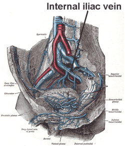
The veins of the right half of the male pelvis.
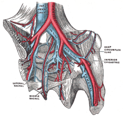
The iliac veins. (Int. iliac visible at center.)
Details
Drains fromPelvic viscera
SourceInternal pudendal vein, middle rectal vein, vesical vein, uterine vein, obturator vein, inferior gluteal vein, superior gluteal vein
Drains toCommon iliac vein
ArteryInternal iliac artery
Identifiers
Latinvena iliaca interna,
vena hypogastrica
TA98A12.3.10.004
TA25024
FMA18884
Anatomical terminology

[edit on Wikidata]

The internal iliac vein (hypogastric vein) begins near the upper part of the greater sciatic foramen, passes upward behind and slightly medial to the internal iliac artery and, at the brim of the pelvis, joins with the external iliac vein to form the common iliac vein.

Structure[edit]

Several veins unite above the greater sciatic foramen to form the internal iliac vein. It does not have the predictable branches of the internal iliac artery but its tributaries drain the same regions.[1] The internal iliac vein emerges from above the level of the greater sciatic notch It runs backwards, upwards and towards the midline to join the external iliac vein in forming the common iliac vein in front of the sacroiliac joint. It usually lies lateral to the internal iliac artery.[2] It is wide and 3 cm long.[3]

Tributaries[edit]

Originating outside the pelvis, its tributaries are the gluteal, internal pudendal and obturator veins. Running from the anterior surface of the sacrum are the lateral sacral veins. Coming from the pelvic plexuses and appropriate to gender are the middle rectal, vesical, prostatic, uterine and vaginal veins.[1][3]

Receives Description
superior gluteal veins
inferior gluteal veins
internal pudendal veins
obturator veins
have their origins outside the pelvis;
lateral sacral veins lie in front of the sacrum
middle hemorrhoidal vein
vesical vein
uterine vein
vaginal veins
originate in venous plexuses connected with the pelvic viscera.

Variation[edit]

On the left, the internal iliac vein lies lateral to the internal iliac artery 73% of the time.[4] On the right, this is 93% of the time.[4]

Function[edit]

The internal iliac veins drain the pelvic organs, sacrum, and coccyx.[2]

Clinical significance[edit]

Ifthrombosis disrupts blood flow in the external iliac systems, the internal iliac tributaries offer a major route of venous return from the femoral system. Damage to internal iliac vein tributaries during surgery can seriously compromise venous drainage and cause swelling of one or both legs.[1]

Additional images[edit]

References[edit]

  1. ^ a b c Delancey, John O.L. (2016). "73, True pelvis, pelvic floor and perineum". In Standring, Susan (ed.). Gray's Anatomy: The Anatomical Basis of Clinical Practice (41st ed.). Elsevier. pp. 1221–1236. ISBN 978-0-7020-6851-5.
  • ^ a b Cramer, Gregory D.; Ro, Chae-Song (January 1, 2014), Cramer, Gregory D.; Darby, Susan A. (eds.), "Chapter 8 - The Sacrum, Sacroiliac Joint, and Coccyx", Clinical Anatomy of the Spine, Spinal Cord, and Ans (Third Edition), Saint Louis: Mosby, pp. 312–339, ISBN 978-0-323-07954-9, retrieved January 28, 2021
  • ^ a b Sinnatamby, Chummy S. (2011). "5". Last's Anatomy: Regional and Applied (12th ed.). Great Britain: Churchill Livingstone Elsevier. p. 309. ISBN 978-0-7020-4839-5. Retrieved March 25, 2018.
  • ^ a b Bleich, April T.; Rahn, David D.; Wieslander, Cecilia K.; Wai, Clifford Y.; Roshanravan, Shayzreen M.; Corton, Marlene M. (December 1, 2007). "Posterior division of the internal iliac artery: Anatomic variations and clinical applications". American Journal of Obstetrics and Gynecology. 197 (6): 658.e1–658.e5. doi:10.1016/j.ajog.2007.08.063. ISSN 0002-9378. PMID 18060970.
  • External links[edit]

    Public domain This article incorporates text in the public domain from page 673 of the 20th edition of Gray's Anatomy (1918)


  • t
  • e

  • Retrieved from "https://en.wikipedia.org/w/index.php?title=Internal_iliac_vein&oldid=1221743330"

    Categories: 
    Wikipedia articles incorporating text from the 20th edition of Gray's Anatomy (1918)
    Veins of the torso
    Angiology
    Cardiovascular system stubs
    Hidden categories: 
    Use American English from January 2019
    All Wikipedia articles written in American English
    Articles with short description
    Short description matches Wikidata
    Use mdy dates from January 2019
    Anatomy NAV infobox with use of other NAV parameters
    Articles with TA98 identifiers
    All stub articles


    This page was last edited on 1 May 2024, at 18:47 (UTC).

    Text is available under the Creative Commons Attribution-ShareAlike License 4.0; additional terms may apply. By using this site, you agree to the Terms of Use and Privacy Policy. Wikipedia® is a registered trademark of the Wikimedia Foundation, Inc., a non-profit organization.


    Privacy policy

    About Wikipedia

    Disclaimers

    Contact Wikipedia

    Code of Conduct

    Developers

    Statistics

    Cookie statement

    Mobile view


    Wikimedia Foundation
    Powered by MediaWiki