Jump to content


Main menu


Navigation  


Main page
Contents
Current events
Random article
About Wikipedia
Contact us
Donate


Contribute  


Help
Learn to edit
Community portal
Recent changes
Upload file


Search  


Create account

Log in


Create account
 Log in


Pages for logged out editors learn more  


Contributions
Talk


Contents


(Top)


1 See also  


2 References  


Microneme


العربية
Français
Galego
Português
Русский
 

Edit links


Article
Talk


Read
Edit
View history


Tools


Actions  


Read
Edit
View history


General  


What links here
Related changes
Upload file
Special pages
Permanent link
Page information
Cite this page
Get shortened URL
Download QR code
Wikidata item


Print/export  


Download as PDF
Printable version


Appearance


From Wikipedia, the free encyclopedia


TachyzoitesofToxoplasma gondii, transmission electron microscopy.[1] Micronemes: mn (click to enlarge)

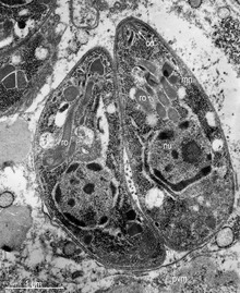
Micronemes are secretory organelles, possessed by parasitic apicomplexans. Micronemes are located on the apical third of the protozoan body. They are surrounded by a typical unit membrane. On electron microscopy they have an electron-dense matrix due to the high protein content. They are specialized secretory organelles important for host-cell invasion and gliding motility.[2]

These organelles secrete several proteins such as the Plasmodium falciparum apical membrane antigen-1, or PfAMA1, and Erythrocyte family antigen, or EBA, family proteins. These proteins specialize in bindingtoerythrocyte surface receptors and facilitating erythrocyte entry. Only by this initial chemical exchange can the parasite enter into the erythrocyte via actin-myosin motor complex.[citation needed]

It has been posited that this organelle works cooperatively with its counterpart organelle, the rhoptry, which also is a secretory organelle. It is possible that, while the microneme initiates erythrocyte-binding, the rhoptry secretes proteins to create the PVM, or the parasitophorous vacuole membrane, in which the parasite can survive and reproduce.[3]

See also[edit]

References[edit]

  1. ^ Rigoulet, Jacques; Hennache, Alain; Lagourette, Pierre; George, Catherine; Longeart, Loïc; Le Net, Jean-Loïc; Dubey, Jitender P. (2014). "Toxoplasmosis in a bar-shouldered dove (Geopelia humeralis) from the Zoo of Clères, France". Parasite. 21: 62. doi:10.1051/parasite/2014062. ISSN 1776-1042. PMC 4236686. PMID 25407506. Open access icon
  • ^ Black, MW; Boothroyd, JC (September 2000). "Lytic cycle of Toxoplasma gondii". Microbiology and Molecular Biology Reviews. 64 (3): 607–23. doi:10.1128/mmbr.64.3.607-623.2000. PMC 99006. PMID 10974128.
  • ^ Lingelbach, K; Joiner, KA (June 1998). "The parasitophorous vacuole membrane surrounding Plasmodium and Toxoplasma: an unusual compartment in infected cells". Journal of Cell Science. 111 ( Pt 11): 1467–75. PMID 9580555.

  • Retrieved from "https://en.wikipedia.org/w/index.php?title=Microneme&oldid=1089968862"

    Category: 
    Parasitology
    Hidden categories: 
    CS1: long volume value
    All articles with unsourced statements
    Articles with unsourced statements from August 2018


    This page was last edited on 26 May 2022, at 16:27 (UTC).

    Text is available under the Creative Commons Attribution-ShareAlike License 4.0; additional terms may apply. By using this site, you agree to the Terms of Use and Privacy Policy. Wikipedia® is a registered trademark of the Wikimedia Foundation, Inc., a non-profit organization.


    Privacy policy

    About Wikipedia

    Disclaimers

    Contact Wikipedia

    Code of Conduct

    Developers

    Statistics

    Cookie statement

    Mobile view


    Wikimedia Foundation
    Powered by MediaWiki