Jump to content


Main menu


Navigation  


Main page
Contents
Current events
Random article
About Wikipedia
Contact us
Donate


Contribute  


Help
Learn to edit
Community portal
Recent changes
Upload file


Search  


Create account

Log in


Create account
 Log in


Pages for logged out editors learn more  


Contributions
Talk


Contents


(Top)


1 Staining  


2 Size and distribution  


3 Functional role  


4 Pathology  


5 References  


6 External links  


Nissl body


العربية
Català
Čeština
Deutsch
Eesti
Español
فارسی
Français
Galego
Italiano
עברית

Oʻzbekcha / ўзбекча
Polski
Português
Română
Русский
Српски / srpski
Українська


Edit links


Article
Talk


Read
Edit
View history


Tools


Actions  


Read
Edit
View history


General  


What links here
Related changes
Upload file
Special pages
Permanent link
Page information
Cite this page
Get shortened URL
Download QR code
Wikidata item


Print/export  


Download as PDF
Printable version


In other projects  


Wikimedia Commons


Appearance


From Wikipedia, the free encyclopedia
 

(Redirected from Nissl substance)

Photomicrograph of Nissl bodies (two are indicated by arrows) in the cytoplasm of motor neurons in the anterior horn of the spinal cord; cresyl violet stain (purple) along with a luxol fast blue stain for myelin. Scale bar = 30 microns (0.03mm).
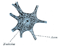
Drawing of a motor neuron from the ventral horn of the medulla spinals of a rabbit. The angular and spindle-shaped Nissl bodies in the cytoplasm are well shown.

Incellular neuroscience, Nissl bodies (also called Nissl granules, Nissl substanceortigroid substance) are discrete granular structures in neurons that consist of rough endoplasmic reticulum, a collection of parallel, membrane-bound cisternae studded with ribosomes on the cytosolic surface of the membranes.[1] Nissl bodies were named after Franz Nissl, a German neuropathologist who invented the staining method bearing his name (Nissl staining).[2][3] The term "Nissl bodies" generally refers to discrete clumps of rough endoplasmic reticulum and free ribosomes in nerve cells. Masses of rough endoplasmic reticulum also occur in some non-neuronal cells, where they are referred to as ergastoplasm, basophilic bodies,[1] or chromophilic substance.[4] While these organelles differ in some ways from Nissl bodies in neurons,[5] large amounts of rough endoplasmic reticulum are generally linked to the copious production of proteins.[1]

Staining[edit]

"Nissl stains" refers to various basic dyes that selectively label negatively charged molecules such as DNA and RNA. Because ribosomes are rich in ribosomal RNA, they are strongly basophilic ("base-loving"). The dense accumulation of membrane-bound and free ribosomes in Nissl bodies results in their intense coloration by Nissl stains, allowing them to be seen with a light microscope.[1]

Size and distribution[edit]

Nissl bodies occur in the somata and dendrites of neurons, though not in the axonoraxon hillock.[6] They vary in size, shape, and intracellular location; they are most conspicuous in the motor neurons of the spinal cord and brainstem, where they appear as large, blocky assemblies.[5] In other neurons, they may be smaller, and in some (such as the granule neurons of the cerebellar cortex) very little rough endoplasmic reticulum is present.[5] The pattern of coloration with Nissl stains once was used to classify neurons.[5] For various reasons, this practice has largely ceased, but specific neuronal types do manifest characteristic types of Nissl bodies.[5]

Functional role[edit]

The functions of Nissl bodies are thought to be the same as those of the rough endoplasmic reticulum in general, primarily the synthesis and segregation of proteins.[1][2] Similar to the ergastoplasm of glandular cells, Nissl bodies are the main site of protein synthesis in the neuronal cytoplasm.[5] The ultrastructure of Nissl bodies suggests they are primarily concerned with the synthesis of proteins for intracellular use.[7]

Pathology[edit]

Nissl bodies show changes under various physiological conditions and in pathological conditions such as axonotmesis, during which they may dissolve and largely disappear (chromatolysis). If the neuron is successful in repairing the damage, the Nissl bodies gradually reappear and return to their characteristic distribution within the cell.[5]

References[edit]

  1. ^ a b c d e Junqueira LC, Carniero J, Kelley RO (1995). Basic Histology. Appleton & Lange. ISBN 0-8385-0567-8.
  • ^ a b Richard H. Thompson (29 March 2000). The Brain: A Neuroscience Primer. Macmillan. pp. 35–. ISBN 978-0-7167-3226-6. Retrieved 4 January 2013.
  • ^ Da Mota Gomes M (2019). "Franz Nissl (1860-1919), noted neuropsychiatrist and neuropathologist, staining the neuron, but not limiting it". Dementia & Neuropsychologia. 13 (3): 352–355. doi:10.1590/1980-57642018dn13-030014. PMC 6753910.
  • ^ Fawcett, Don W. (1966). The Cell: Its Organelles and Inclusions. W.B. Saunders Company. ISBN 0-7216-3585-7.
  • ^ a b c d e f g Peters A, Palay SL, Webster Hd (1991). The Fine Structure of the Nervous System. Oxford University Press. ISBN 0-19-506571-9.
  • ^ Wolfgang Kühnel (2003). Color Atlas of Cytology, Histology, and Microscopic Anatomy. Thieme. pp. 182–. ISBN 978-3-13-562404-4. Retrieved 4 January 2013.
  • ^ T. Herdegen; J. Delgado-Garcia (25 May 2005). Brain Damage and Repair: From Molecular Research to Clinical Therapy. Springer. pp. 37–. ISBN 978-1-4020-1892-3. Retrieved 4 January 2013.
  • External links[edit]


    Retrieved from "https://en.wikipedia.org/w/index.php?title=Nissl_body&oldid=1171739308"

    Category: 
    Nervous tissue cells
    Hidden categories: 
    Articles with short description
    Short description is different from Wikidata
    Commons category link is on Wikidata


    This page was last edited on 22 August 2023, at 23:06 (UTC).

    Text is available under the Creative Commons Attribution-ShareAlike License 4.0; additional terms may apply. By using this site, you agree to the Terms of Use and Privacy Policy. Wikipedia® is a registered trademark of the Wikimedia Foundation, Inc., a non-profit organization.


    Privacy policy

    About Wikipedia

    Disclaimers

    Contact Wikipedia

    Code of Conduct

    Developers

    Statistics

    Cookie statement

    Mobile view


    Wikimedia Foundation
    Powered by MediaWiki