Jump to content


Main menu


Navigation  


Main page
Contents
Current events
Random article
About Wikipedia
Contact us
Donate


Contribute  


Help
Learn to edit
Community portal
Recent changes
Upload file


Search  


Create account

Log in


Create account
 Log in


Pages for logged out editors learn more  


Contributions
Talk


Contents


(Top)


1 Pseudopupil analysis technique  


2 References  


Pseudopupil


العربية
Deutsch
Bahasa Indonesia
Українська
 

Edit links


Article
Talk


Read
Edit
View history


Tools


Actions  


Read
Edit
View history


General  


What links here
Related changes
Upload file
Special pages
Permanent link
Page information
Cite this page
Get shortened URL
Download QR code
Wikidata item


Print/export  


Download as PDF
Printable version


In other projects  


Wikimedia Commons


Appearance


From Wikipedia, the free encyclopedia


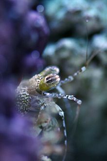
The head of a mantis showing the black pseudopupil in its compound eyes
The eye of a mantis shrimp has three regions, each with its own pseudopupil.

In the compound eyeofinvertebrates such as insects and crustaceans, the pseudopupil appears as a dark spot which moves across the eye as the animal is rotated.[1] This occurs because the ommatidia that one observes "head-on" (along their optical axes) absorb the incident light, while those to one side reflect it.[2] The pseudopupil therefore reveals which ommatidia are aligned with the axis along which the observer is viewing.[2]

Pseudopupil analysis technique[edit]

The pseudopupil analysis technique is used to study neurodegeneration in insects like Drosophila. An adult Drosophila eye consists of nearly 800 unit ommatidia which are repeated in a symmetrical pattern. Each ommatidium contains 8 photoreceptor cells, each of which forms a rhabdomere (rhabdomeres 7 and 8 overlap vertically; therefore, only rhabdomere 7 is visible externally). Neurodegeneration leads to loss or degradation of photoreceptors.[3] By visualizing and counting the intact rhabdomeres, degradation level can be measured. Thus, analyzing the pseudopupil can permit empirical study of neurodegeneration.

References[edit]

  1. ^ M. F. Land; G. Gibson; J. Horwood; J. Zeil (1999). "Fundamental differences in the optical structure of the eyes of nocturnal and diurnal mosquitoes" (PDF). Journal of Comparative Physiology A. 185 (1): 91–103. doi:10.1007/s003590050369. S2CID 9114187. Archived from the original (PDF) on 2016-03-04. Retrieved 2008-07-27.
  • ^ a b Jochen Zeil & Maha M. Al-Mutairi (1996). "Variations in the optical properties of the compound eyes of Uca lactea annulipes" (PDF). The Journal of Experimental Biology. 199 (7): 1569–1577. PMID 9319471.
  • ^ Song, Wan; Smith, Marianne R.; Syed, Adeela; Lukacsovich, Tamas; Barbaro, Brett A.; Purcell, Judith; Bornemann, Doug J.; Burke, John; Marsh, J. Lawrence (2013). Morphometric analysis of Huntington's disease neurodegeneration in Drosophila. Methods in Molecular Biology. Vol. 1017. pp. 41–57. doi:10.1007/978-1-62703-438-8_3. ISBN 978-1-62703-437-1. ISSN 1940-6029. PMID 23719906.
  • t
  • e

  • Retrieved from "https://en.wikipedia.org/w/index.php?title=Pseudopupil&oldid=1147290673"

    Categories: 
    Eye
    Arthropod anatomy
    Eye stubs
    Hidden categories: 
    Articles with short description
    Short description matches Wikidata
    Commons category link is on Wikidata
    All stub articles


    This page was last edited on 30 March 2023, at 00:09 (UTC).

    Text is available under the Creative Commons Attribution-ShareAlike License 4.0; additional terms may apply. By using this site, you agree to the Terms of Use and Privacy Policy. Wikipedia® is a registered trademark of the Wikimedia Foundation, Inc., a non-profit organization.


    Privacy policy

    About Wikipedia

    Disclaimers

    Contact Wikipedia

    Code of Conduct

    Developers

    Statistics

    Cookie statement

    Mobile view


    Wikimedia Foundation
    Powered by MediaWiki