Jump to content


Main menu


Navigation  


Main page
Contents
Current events
Random article
About Wikipedia
Contact us
Donate


Contribute  


Help
Learn to edit
Community portal
Recent changes
Upload file


Search  


Create account

Log in


Create account
 Log in


Pages for logged out editors learn more  


Contributions
Talk


Contents


(Top)


1 See also  


2 External links  


3 Notes  


Serous demilune


العربية


Edit links


Article
Talk


Read
Edit
View history


Tools


Actions  


Read
Edit
View history


General  


What links here
Related changes
Upload file
Special pages
Permanent link
Page information
Cite this page
Get shortened URL
Download QR code
Wikidata item


Print/export  


Download as PDF
Printable version


Appearance


From Wikipedia, the free encyclopedia


Serous demilune
Section of submandibular gland of kitten. Duct semidiagrammatic. (Demilune labeled at center bottom.)
Identifiers
THH2.00.02.0.03062
Anatomical terminology

[edit on Wikidata]

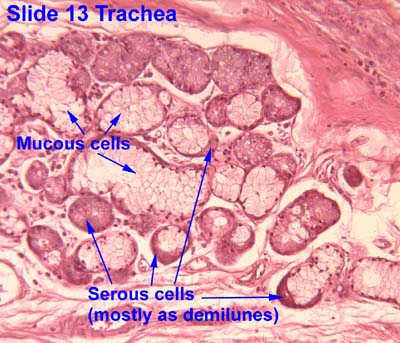
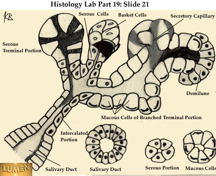
Serous demilunes, also known as Crescents of GiannuzziorDemilunes of Heidenhain, are cellular formations in the shape of a half-moon (hence the name "demilune") on the mixed submandibular and sublingual salivary glands.[1]

Serous demilunes are the serous cells at the distal end of mucous acini, the secretory endpieces of certain salivary glands.[1] These demilune cells secrete the proteins that contain the enzyme lysozyme, which degrades the cell walls of bacteria. In this way, lysozyme confers antimicrobial activity to mucus.

The serous demilune is an artifact from traditional methods of preparing samples. Samples are traditionally preserved and fixed in formaldehyde. When samples were preserved by quick-freezing in liquid nitrogen and then fixed with osmium tetraoxide in acetone, no demilunes were found. Examination showed that the serous cells and mucosal cells were aligned in the acinus. The traditional preparation caused mucous cells to swell during fixation which results in the serous cells being popped out of their alignment. After sectioning the serous cells resembled the common demilune shape, and were so named.[2]

When the gland has this demilunar structure, it has mucoserous acini producing both serous and mucous secretions.

Slice of submandibular gland. H&E staining, the structure of the red stained cytoplasm on the left is the serous demilunes.

See also

[edit]
[edit]

Notes

[edit]
  1. ^ a b Amano, O; Mizobe, K; Bando, Y; Sakiyama, K (31 October 2012). "Anatomy and histology of rodent and human major salivary glands: -overview of the Japan salivary gland society-sponsored workshop-". Acta Histochemica et Cytochemica. 45 (5): 241–50. doi:10.1267/ahc.12013. PMC 3496860. PMID 23209333.
  • ^ Ross, Michael (2003). Histology: A Text and Atlas. Lippincott Williams & Wilkins. pp. 434–474. ISBN 0-7817-5124-1.

  • Retrieved from "https://en.wikipedia.org/w/index.php?title=Serous_demilune&oldid=1235814422"

    Category: 
    Glands


    This page was last edited on 21 July 2024, at 10:49 (UTC).

    Text is available under the Creative Commons Attribution-ShareAlike License 4.0; additional terms may apply. By using this site, you agree to the Terms of Use and Privacy Policy. Wikipedia® is a registered trademark of the Wikimedia Foundation, Inc., a non-profit organization.


    Privacy policy

    About Wikipedia

    Disclaimers

    Contact Wikipedia

    Code of Conduct

    Developers

    Statistics

    Cookie statement

    Mobile view


    Wikimedia Foundation
    Powered by MediaWiki