
| Superior orbital fissure | |
|---|---|

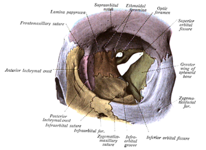
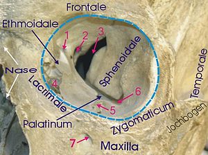
Orbit seen from the front, with bones labeled in different colors, and superior orbital fissure at center as an "hour-glass" formation.
| |

1Foramen ethmoidale, 2 Canalis opticus, 3 Fissura orbitalis superior, 4 Fossa sacci lacrimalis, 5 Sulcus infraorbitalis, 6 Fissura orbitalis inferior, 7 Foramen infraorbitale
| |
| Details | |
| Part of | Sphenoid bone |
| System | Skeletal |
| Identifiers | |
| Latin | fissura orbitalis superior |
| TA98 | A02.1.05.023 A02.1.00.083 |
| TA2 | 488 |
| FMA | 54799 |
| Anatomical terms of bone | |
The superior orbital fissure is a foramen or cleft of the skull between the lesser and greater wings of the sphenoid bone. It gives passage to multiple structures, including the oculomotor nerve, trochlear nerve, ophthalmic nerve, abducens nerve, ophthalmic veins, and sympathetic fibres from the cavernous plexus.

The superior orbital fissure is usually 22 mm wide in adults,[1] and is much larger medially. Its boundaries are formed by the (caudal surface of the) lesser wing of the sphenoid bone, and (medial border of the) greater wing of the sphenoid bone.[2]
The superior orbital fissure is traversed by the following structures:
The superior orbital fissure is divided into 3 parts from lateral to medial:[citation needed]
Multiple anatomical structures pass through the fissure, and can be damaged in orbital trauma, particularly blowout fractures through the floor of the orbit into the maxillary sinus.[citation needed]
The abducens nerve is most likely to show signs of damage first, with the most common complaints retro-orbital pain and the involvement of cranial nerves III, IV, V1, and VI without other neurological signs or symptoms. This presentation indicates either compression of structures in the superior orbital fissure or the cavernous sinus.[citation needed]
Superior orbital fissure syndrome, also known as Rochon-Duvigneaud's syndrome,[4][5] is a neurological disorder that results if the superior orbital fissure is fractured. Involvement of the cranial nerves that pass through the superior orbital fissure may lead to diplopia, paralysis of extraocular muscles, exophthalmos, and ptosis. Blindness or loss of vision indicates involvement of the orbital apex, which is more serious, requiring urgent surgical intervention. Typically, if blindness is present with superior orbital syndrome, it is called orbital apex syndrome.[citation needed]
{{cite book}}: CS1 maint: date and year (link)
|
Neurocranium of the skull
| |||||||||||
|---|---|---|---|---|---|---|---|---|---|---|---|
| Occipital |
| ||||||||||
| Parietal |
| ||||||||||
| Frontal |
| ||||||||||
| Temporal |
| ||||||||||
| Sphenoid |
| ||||||||||
| Ethmoid |
| ||||||||||
|
| |||||||||
|---|---|---|---|---|---|---|---|---|---|
| Anterior cranial fossa |
| ||||||||
| Middle cranial fossa |
| ||||||||
| Posterior cranial fossa |
| ||||||||
| Orbit |
| ||||||||
| Pterygopalatine fossa |
| ||||||||
| |||||||||
| Other |
| ||||||||