Jump to content


Main menu


Navigation  


Main page
Contents
Current events
Random article
About Wikipedia
Contact us
Donate


Contribute  


Help
Learn to edit
Community portal
Recent changes
Upload file


Search  


Create account

Log in


Create account
 Log in


Pages for logged out editors learn more  


Contributions
Talk


Contents


(Top)


1 Clinical significance  


2 Other animals  


3 Additional images  


4 See also  


5 References  


5.1  Sources  


6 External links  


Urogenital sinus


فارسی

Polski
Português
Română
Русский
 

Edit links


Article
Talk


Read
Edit
View history


Tools


Actions  


Read
Edit
View history


General  


What links here
Related changes
Upload file
Special pages
Permanent link
Page information
Cite this page
Get shortened URL
Download QR code
Wikidata item


Print/export  


Download as PDF
Printable version


Appearance


From Wikipedia, the free encyclopedia


Urogenital sinus
Urogenital sinus of female human embryo of eight and a half to nine weeks old. (Urogenital sinus labeled at bottom.)
Stages in the development of the external sexual organs in the male and female. ("Opening of urogenital sinus" labeled in diagram D.)
Details
Carnegie stage15
PrecursorCloaca
Gives rise toUrethra, bladder, vagina, vulval vestibule, Bartholin's glands, Skene's glands, prostate, bulbourethral glands
Identifiers
Latinsinus urogenitalis definitivus
TEsinus_by_E5.7.3.1.0.0.1 E5.7.3.1.0.0.1
Anatomical terminology

[edit on Wikidata]

The urogenital sinus is a body part of a human or other placental only present in the development of the urinary and reproductive organs. It is the ventral part of the cloaca, formed after the cloaca separates from the anal canal during the fourth to seventh weeks of development.[1]

In males, the UG sinus is divided into three regions: upper, pelvic, and phallic. The upper part gives rise to the urinary bladder and the pelvic part gives rise to the prostatic and membranous parts of the urethra,[1] the prostate and the bulbourethral glands (Cowper's). The phallic portion gives rise to the spongy (bulbar) part of the urethra and the urethral glands (Littré's).

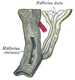
In females, the pelvic part of the UG sinus gives rise to the sinovaginal bulbs, structures that will eventually form the inferior two thirds of the vagina. This process begins when the lower tip of the paramesonephric ducts, the structures that will eventually form the uterus and vaginal fornices, come in contact with the UG sinus. Shortly afterwards, the sinovaginal bulbs form as two solid evaginations of the UG sinus. Cells in these bulbs divide to form a solid vaginal plate, which extends and then canalizes (hollows) to form the inferior portion of the vagina.[2] The female urogenital sinus also gives rise to the urethra, vestibule, Skene's glands and Bartholin's glands.

Clinical significance[edit]

Aurogenital sinus anomaly is also a rare birth defectinwomen where the urethra and vagina both open into a common channel.[3][4]

Apersistent cloaca is a disorder where the rectum, vagina, and urinary tract meet and fuse, creating a cloaca, a single common channel.[5]

Other animals[edit]

In many mammals (excluding primates),[6] the urogenital sinus refers to the sinus in which the openings to the female's urethra and vagina are found.[6][7][8][9] However, this term may also apply to some male mammals. The urogenital sinus of non-primates is homologous to the vulval vestibule of primates.

Additional images[edit]

See also[edit]

References[edit]

  1. ^ a b Sadler & Langman 2010, pp. 243–244.
  • ^ Sadler & Langman 2010, pp. 253.
  • ^ Callahan, Gerald N. (2009). Between XX and XY : intersexuality and the myth of two sexes. Chicago, Ill.: Chicago Review Press. p. 182. ISBN 978-1-56976-289-9. OCLC 436089205.
  • ^ "Urogenital Sinus". Children's Hospital of Orange County. 2023-02-23. Retrieved 2023-10-07.
  • ^ Jenkins D, Bitner-Glindzicz M, Thomasson L, et al. (2007), "Mutational analyses of UPIIIA, SHH, EFNB2 and HNF1β in persistent cloaca and associated kidney malformations", J Pediatr Urol, 3 (1): 2–9, doi:10.1016/j.jpurol.2006.03.002, PMC 1864944, PMID 17476318.
  • ^ a b Schatten, Heide; Constantinescu, Gheorghe M. (2007-08-14). Comparative Reproductive Biology. John Wiley & Sons. ISBN 978-0-8138-1554-1.
  • ^ Armati, Patricia J.; Dickman, Chris R.; Hume, Ian D. (2006-08-17). Marsupials. Cambridge University Press. ISBN 978-1-139-45742-2.
  • ^ Grant, Tom (1995). The Platypus: A Unique Mammal. UNSW Press. ISBN 978-0-86840-143-0.
  • ^ Tyndale-Biscoe, C. Hugh; Renfree, Marilyn (1987-01-30). Reproductive Physiology of Marsupials. Cambridge University Press. ISBN 978-0-521-33792-2.
  • Sources[edit]

    External links[edit]


    Retrieved from "https://en.wikipedia.org/w/index.php?title=Urogenital_sinus&oldid=1234123364"

    Category: 
    Embryology of urogenital system
    Hidden categories: 
    Articles with short description
    Short description is different from Wikidata


    This page was last edited on 12 July 2024, at 18:10 (UTC).

    Text is available under the Creative Commons Attribution-ShareAlike License 4.0; additional terms may apply. By using this site, you agree to the Terms of Use and Privacy Policy. Wikipedia® is a registered trademark of the Wikimedia Foundation, Inc., a non-profit organization.


    Privacy policy

    About Wikipedia

    Disclaimers

    Contact Wikipedia

    Code of Conduct

    Developers

    Statistics

    Cookie statement

    Mobile view


    Wikimedia Foundation
    Powered by MediaWiki