Jump to content


Main menu


Navigation  


Main page
Contents
Current events
Random article
About Wikipedia
Contact us
Donate


Contribute  


Help
Learn to edit
Community portal
Recent changes
Upload file


Search  


Create account

Log in


Create account
 Log in


Pages for logged out editors learn more  


Contributions
Talk


Contents


(Top)


1 Common methods  


1.1  Frogging and tadpoling  


2 List of viability assay methods  


3 See also  


4 References  


5 Further reading  


Viability assay


Eesti
 

Edit links


Article
Talk


Read
Edit
View history


Tools


Actions  


Read
Edit
View history


General  


What links here
Related changes
Upload file
Special pages
Permanent link
Page information
Cite this page
Get shortened URL
Download QR code
Wikidata item


Print/export  


Download as PDF
Printable version


Appearance


From Wikipedia, the free encyclopedia


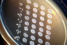
This plated viability assay measures various yeast viability though a method called "frogging". The research is completed through drop-inoculation techniques. Research has since been conducted on "tadpoling", which is a variation of "frogging" that is completed by keeping the test cells diluted in liquid throughout their examination.[1]

Aviability assay is an assay that is created to determine the ability of organs, cellsortissues to maintain or recover a state of survival.[2] Viability can be distinguished from the all-or-nothing states of life and death by the use of a quantifiable index that ranges between the integers of 0 and 1 or, if more easily understood, the range of 0% and 100%.[3] Viability can be observed through the physical properties of cells, tissues, and organs. Some of these include mechanical activity, motility, such as with spermatozoa and granulocytes, the contraction of muscle tissue or cells, mitotic activity in cellular functions, and more.[3] Viability assays provide a more precise basis for measurement of an organism's level of vitality.

Viability assays can lead to more findings than the difference of living versus nonliving. These techniques can be used to assess the success of cell culture techniques, cryopreservation techniques, the toxicity of substances, or the effectiveness of substances in mitigating effects of toxic substances.[4]

Common methods[edit]

Though simple visual techniques of observing viability can be useful, it can be difficult to thoroughly measure an organism's/part of an organism's viability merely using the observation of physical properties. However, there are a variety of common protocols utilized for further observation of viability using assays.

Flow cytometry using 7-Aminoactinomycin D (7-AAD), wherein a lower signal indicates viable cells. Therefore, this case shows good viability (viability of the cells in flow cytometry should be around 95% but not less than 90%.[8]).

As with many kinds of viability assays, quantitative measures of physiological function do not indicate whether damage repair and recovery is possible.[12] An assay of the ability of a cell line to adhere and divide may be more indicative of incipient damage than membrane integrity.[13]

Frogging and tadpoling[edit]

"Frogging" is a type of viability assay method that utilizes an agar plate for its environment and consists of plating serial dilutions by pinning them after they have been diluted in liquid. Some of its limitations include that it does not account for total viability and it is not particularly sensitive to low-viability assays; however, it is known for its quick pace.[1] "Tadpoling", which is a method practiced after the development of "frogging", is similar to the "frogging" method, but its test cells are diluted in liquid and then kept in liquid through the examination process. The "tadpoling" method can be used to measure culture viability accurately, which is what depicts its main separation from "frogging".[1]

List of viability assay methods[edit]

See also[edit]

References[edit]

  1. ^ a b c Welch AZ, Koshland DE (2013). "A simple colony-formation assay in liquid medium, termed 'tadpoling', provides a sensitive measure of Saccharomyces cerevisiae culture viability". Yeast. 30 (12): 501–509. doi:10.1002/yea.2989. ISSN 1097-0061. PMID 24185677. S2CID 21664332.
  • ^ Gavanji S, Bakhtari A, Famurewa AC, Othman EM (January 2023). "Cytotoxic Activity of Herbal Medicines as Assessed in Vitro: A Review". Chemistry & Biodiversity. 20 (2): 3–27. doi:10.1002/cbdv.202201098. PMID 36595710. S2CID 255473013.
  • ^ a b Pegg DE (1989-06-01). "Viability assays for preserved cells, tissues, and organs". Cryobiology. 26 (3): 212–231. doi:10.1016/0011-2240(89)90016-3. ISSN 0011-2240. PMID 2743785.
  • ^ "Overview of Probes for Cell Viability, Cell Proliferation and Live-Cell Function—Section 15.1 - US". www.thermofisher.com. Retrieved 2020-03-04.
  • ^ a b c d Niles AL, Moravec RA, Worzella TJ, Evans NJ, Riss TL (2013-03-04). "High-Throughput Screening Assays for the Assessment of Cytotoxicity". In Steinberg P (ed.). High-Throughput Screening Methods in Toxicity Testing. John Wiley & Sons, Inc. pp. 107–127. doi:10.1002/9781118538203.ch5. ISBN 978-1-118-53820-3.
  • ^ Lindner B, Seydel U (January 1983). "Mass spectrometric analysis of drug-induced changes in Na+ and K+ contents of single bacterial cells". Journal of General Microbiology. 129 (1): 51–5. doi:10.1099/00221287-129-1-51. PMID 6339677.
  • ^ Pichugin Y, Fahy GM, Morin R (April 2006). "Cryopreservation of rat hippocampal slices by vitrification". Cryobiology. 52 (2): 228–40. doi:10.1016/j.cryobiol.2005.11.006. PMID 16403489.
  • ^ "Flow cytometry (FACS) staining protocol (Cell surface staining)". Yale School of Medicine - Yale Flow Cytometry. Retrieved 2023-10-17.
  • ^ Henkelman S, Lagerberg JW, Graaff R, Rakhorst G, Van Oeveren W (November 2010). "The effects of cryopreservation on red blood cell rheologic properties". Transfusion. 50 (11): 2393–401. doi:10.1111/j.1537-2995.2010.02730.x. PMID 20561300. S2CID 22548108.
  • ^ Southard JH (June 1989). "Viability assays in organ preservation". Cryobiology. 26 (3): 232–8. doi:10.1016/0011-2240(89)90017-5. PMID 2663353.
  • ^ Davey HM (August 2011). "Life, death, and in-between: meanings and methods in microbiology". Applied and Environmental Microbiology. 77 (16): 5571–6. Bibcode:2011ApEnM..77.5571D. doi:10.1128/AEM.00744-11. PMC 3165249. PMID 21705550.
  • ^ Crutchfield A, Diller K, Brand J (1999-02-01). "Cryopreservation of Chlamydomonas reinhardtii (Chlorophyta)". European Journal of Phycology. 34 (1): 43–52. Bibcode:1999EJPhy..34...43C. doi:10.1080/09670269910001736072.
  • ^ Wusteman MC, Pegg DE, Robinson MP, Wang LH, Fitch P (February 2002). "Vitrification media: toxicity, permeability, and dielectric properties". Cryobiology. 44 (1): 24–37. doi:10.1016/S0011-2240(02)00002-0. PMID 12061845.
  • Further reading[edit]

  • Riss TL, Moravec RA, Niles AL, Duellman S, Benink HA, Worzella TJ, et al. (2004). "Cell Viability Assays". In Sittampalam GS, Grossman A, Brimacombe K, et al. (eds.). Assay Guidance Manual. Bethesda (MD): Eli Lilly & Company and the National Center for Advancing Translational Sciences. PMID 23805433.

  • Retrieved from "https://en.wikipedia.org/w/index.php?title=Viability_assay&oldid=1204855089"

    Categories: 
    Cell biology
    Cell culture techniques
    Molecular biology techniques
    Microbiology techniques
    Toxicology tests
    Hidden categories: 
    Articles with short description
    Short description is different from Wikidata


    This page was last edited on 8 February 2024, at 05:21 (UTC).

    Text is available under the Creative Commons Attribution-ShareAlike License 4.0; additional terms may apply. By using this site, you agree to the Terms of Use and Privacy Policy. Wikipedia® is a registered trademark of the Wikimedia Foundation, Inc., a non-profit organization.


    Privacy policy

    About Wikipedia

    Disclaimers

    Contact Wikipedia

    Code of Conduct

    Developers

    Statistics

    Cookie statement

    Mobile view


    Wikimedia Foundation
    Powered by MediaWiki