Jump to content


Main menu


Navigation  


Main page
Contents
Current events
Random article
About Wikipedia
Contact us
Donate


Contribute  


Help
Learn to edit
Community portal
Recent changes
Upload file


Search  


Create account

Log in


Create account
 Log in


Pages for logged out editors learn more  


Contributions
Talk


Contents


(Top)


1 Structure  


1.1  Muscle attachments  


2 Injuries  


3 Additional images  


4 Other animals  


5 See also  


6 References  


Cuneiform bones


العربية
Azərbaycanca
Башҡортса
Bosanski
Brezhoneg
Català
Чӑвашла
Deutsch
Español
Euskara
فارسی
Français

Italiano
Македонски
Nederlands
Polski
Português
Русский
Svenska
Türkçe
Українська


Edit links


Article
Talk


Read
Edit
View history


Tools


Actions  


Read
Edit
View history


General  


What links here
Related changes
Upload file
Special pages
Permanent link
Page information
Cite this page
Get shortened URL
Download QR code
Wikidata item


Print/export  


Download as PDF
Printable version


In other projects  


Wikimedia Commons


Appearance


From Wikipedia, the free encyclopedia


Cuneiform bones; Cuneiform
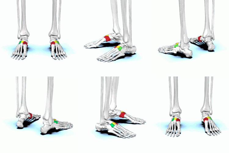
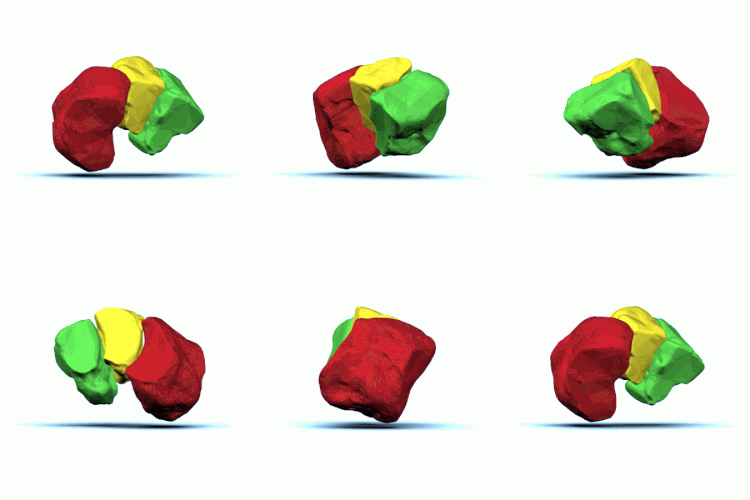
Red=medial; yellow=intermediate; green=lateral
Cuneiform bones of the left foot
Details
Identifiers
Latinos cuneiformis
pl. ossa cuneiformia
FMA24517
Anatomical terms of bone

[edit on Wikidata]

There are three cuneiform ("wedge-shaped") bones in the human foot:

They are located between the navicular bone and the first, second and third metatarsal bones and are medial to the cuboid bone.[1]

Structure[edit]

There are three cuneiform bones:

  1. The medial cuneiform (also known as first cuneiform) is the largest of the cuneiforms. It is situated at the medial side of the foot, anterior to the navicular bone and posterior to the base of the first metatarsal. Lateral to it is the intermediate cuneiform. It articulates with four bones: the navicular, second cuneiform, and first and second metatarsals. The tibialis anterior and fibularis longus muscle inserts at the medial cuneiform bone.[2]
  2. The intermediate cuneiform (second cuneiformormiddle cuneiform) is shaped like a wedge, the thin end pointing downwards. The intermediate cuneiform is situated between the other two cuneiform bones (the medial and lateral cuneiforms), and articulates with the navicular posteriorly, the second metatarsal anteriorly and with the other cuneiforms on either side.
  3. The lateral cuneiform (also known as third cuneiformorexternal cuneiform) intermediate in size between the other two cuneiform bones, is also wedge-shaped, the base being uppermost. It occupies the center of the front row of the tarsal bones, between the intermediate cuneiform medially, the cuboid laterally, the navicular posteriorly and the third metatarsal in front. The tibialis posterior inserts at the lateral cuneiform, while the flexor hallucis brevis originates from it.[2]

Muscle attachments[edit]

Muscle Direction Attachment[2]
Tibialis anterior Insertion Medial cuneiform
Fibularis longus Insertion Medial cuneiform
Tibialis posterior Insertion Medial cuneiform
Flexor hallucis brevis Origin Lateral cuneiform

Injuries[edit]

Additional images[edit]

Other animals[edit]

See also[edit]

References[edit]

  1. ^ Bojsen-Møller, Finn; Simonsen, Erik B.; Tranum-Jensen, Jørgen (2001). Bevægeapparatets anatomi [Anatomy of the Locomotive Apparatus] (in Danish) (12th ed.). p. 245. ISBN 978-87-628-0307-7.
  • ^ a b c Bojsen-Møller, Finn; Simonsen, Erik B.; Tranum-Jensen, Jørgen (2001). Bevægeapparatets anatomi [Anatomy of the Locomotive Apparatus] (in Danish) (12th ed.). pp. 364–367. ISBN 978-87-628-0307-7.
  • ^ TheFreeDictionary > Lisfranc's fracture Citing: Mosby's Medical Dictionary, 8th edition. 2009
  • ^ Mabry LM, Patti TN, Ross MD, Bleakley CM, Gisselman AS (July 2021). "Isolated Medial Cuneiform Fractures: A Systematic Search and Qualitative Analysis of Case Studies". J Am Podiatr Med Assoc. 111 (4): 1–9. doi:10.7547/20-047. PMID 34478529. S2CID 225705519.{{cite journal}}: CS1 maint: multiple names: authors list (link)

  • Retrieved from "https://en.wikipedia.org/w/index.php?title=Cuneiform_bones&oldid=1188620915"

    Categories: 
    Bones of the foot
    Tarsal bones
    Hidden categories: 
    CS1 Danish-language sources (da)
    CS1 maint: multiple names: authors list
    Articles with short description
    Short description is different from Wikidata
    Articles to be expanded from April 2015
    All articles to be expanded
    Articles using small message boxes
    Commons category link is on Wikidata


    This page was last edited on 6 December 2023, at 16:13 (UTC).

    Text is available under the Creative Commons Attribution-ShareAlike License 4.0; additional terms may apply. By using this site, you agree to the Terms of Use and Privacy Policy. Wikipedia® is a registered trademark of the Wikimedia Foundation, Inc., a non-profit organization.


    Privacy policy

    About Wikipedia

    Disclaimers

    Contact Wikipedia

    Code of Conduct

    Developers

    Statistics

    Cookie statement

    Mobile view


    Wikimedia Foundation
    Powered by MediaWiki