Jump to content


Main menu


Navigation  


Main page
Contents
Current events
Random article
About Wikipedia
Contact us
Donate


Contribute  


Help
Learn to edit
Community portal
Recent changes
Upload file


Search  


Create account

Log in


Create account
 Log in


Pages for logged out editors learn more  


Contributions
Talk


Contents


(Top)


1 Anatomy  


2 Histology  


3 Clinical significance  


4 See also  


5 References  


6 External links  


Epoophoron


العربية
Bosanski
Esperanto
Italiano
Polski
Română
Русский
Українська
 

Edit links


Article
Talk


Read
Edit
View history


Tools


Actions  


Read
Edit
View history


General  


What links here
Related changes
Upload file
Special pages
Permanent link
Page information
Cite this page
Get shortened URL
Download QR code
Wikidata item


Print/export  


Download as PDF
Printable version


Appearance


From Wikipedia, the free encyclopedia


Epoophoron
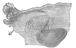
Broad ligament of adult, showing epoöphoron. (From Farre, after Kobelt.) a, a. Epoöphoron formed from the upper part of the mesonephric body. b. Remains of the uppermost tubes sometimes forming appendices. c. Middle set of tubes. d. Some lower atrophied tubes. e. Atrophied remains of the mesonephric duct. f. The terminal bulb or hydatid. h. The uterine tube, originally the duct of Müller. i. Appendix attached to the extremity. l. The ovary.
Uterus and right broad ligament, seen from behind. The epoophoron is visible in upper right
Details
PrecursorMesonephric ducts[1]
Identifiers
TA98A09.1.05.001
TA23540
FMA18691
Anatomical terminology

[edit on Wikidata]

The epoophoronorepoöphoron (also called organ of Rosenmüller[2][3] or the parovarium; pl.: epoophora) is a remnant of the mesonephric duct that can be found next to the ovary and fallopian tube.

Anatomy[edit]

It may contain 10–15 transverse small ducts or tubules that lead to the Gartner's duct (also longitudinal duct of epoophoron) that represents the caudal remnant of the mesonephric ducts and passes through the broad ligament and the lateral wall of the cervix and vagina.

The epoophoron is a homologue to the epididymis in the male.

While the epoophoron is located in the lateral portion of the mesosalpinx and mesovarium, the paroophoron (residual remnant of that part of the mesonephric duct that forms the paradidymis in the male) lies more medially in the mesosalpinx.

Histology[edit]

It has a unique histological profile.[4][5]

Clinical significance[edit]

Clinically the organ may give rise to a local paraovarian cystoradenoma.

See also[edit]

References[edit]

  1. ^ Netter, Frank H.; Cochard, Larry R. (2002). Netter's Atlas of human embryology. Teterboro, N.J: Icon Learning Systems. p. 173. ISBN 0-914168-99-1.
  • ^ synd/2662atWho Named It?
  • ^ J. C. Rosenmüller. De ovariis embryonum et foetuum humanorum. 1802.
  • ^ Woolnough E, Russo L, Khan MS, Heatley MK (2000). "An immunohistochemical study of the rete ovarii and epoophoron". Pathology. 32 (2): 77–83. doi:10.1080/003130200104277. PMID 10840824. S2CID 42714478.
  • ^ Russo L, Woolmough E, Heatley MK (2000). "Structural and cell surface antigen expression in the rete ovarii and epoophoron differs from that in the Fallopian tube and in endometriosis". Histopathology. 37 (1): 64–9. doi:10.1046/j.1365-2559.2000.00938.x. PMID 10931220. S2CID 35199140.
  • External links[edit]


    Retrieved from "https://en.wikipedia.org/w/index.php?title=Epoophoron&oldid=1222354553"

    Category: 
    Mammal female reproductive system
    Hidden categories: 
    Articles with short description
    Short description is different from Wikidata
    Articles with TA98 identifiers


    This page was last edited on 5 May 2024, at 15:04 (UTC).

    Text is available under the Creative Commons Attribution-ShareAlike License 4.0; additional terms may apply. By using this site, you agree to the Terms of Use and Privacy Policy. Wikipedia® is a registered trademark of the Wikimedia Foundation, Inc., a non-profit organization.


    Privacy policy

    About Wikipedia

    Disclaimers

    Contact Wikipedia

    Code of Conduct

    Developers

    Statistics

    Cookie statement

    Mobile view


    Wikimedia Foundation
    Powered by MediaWiki