Oligodendrocytes (from Greek 'cells with a few branches'), also known as oligodendroglia, are a type of neuroglia whose main functions are to provide support and insulation to axons within the central nervous system (CNS) of jawed vertebrates. Their function is similar to that of Schwann cells, which perform the same task in the peripheral nervous system (PNS). Oligodendrocytes accomplish this by forming the myelin sheath around axons.[1] Unlike Schwann cells, a single oligodendrocyte can extend its processes to cover around 50 axons,[2] with each axon being wrapped in approximately 1 μm of myelin sheath. Furthermore, an oligodendrocyte can provide myelin segments for multiple adjacent axons.[1]
Oligodendrocytes are exclusively found in the CNS, which comprises the brain and spinal cord. Initially, it was originally thought that these cells were produced in the ventral neural tube, the embryonic precursor to the CNS. However, recent research suggests that oligodendrocytes originate from the ventral ventricular zone of the embryonic spinal cord, with some potential concentrations in the forebrain.[3] Notably, oligodendrocytes are the last type of cell to be generated in the CNS.[4] Oligodendrocytes were discovered by Pío del Río Hortega.[5][6]
Most oligodendrocytes develop during embryogenesis and early postnatal life from restricted periventricular germinal regions.[11] Oligodendrocyte formation in the adult brain is associated with glial-restricted progenitor cells, known as oligodendrocyte progenitor cells (OPCs).[12]Subventricular zone cells migrate away from germinal[12] zones to populate both developing white and gray matter, where they differentiate and mature into myelin-forming oligodendrocytes.[13] However, it is not clear whether all oligodendrocyte progenitors undergo this sequence of events.[citation needed]
Between midgestation and term birth in human cerebral white matter, three successive stages of the classic human oligodendrocyte lineage are found: OPCs, immature oligodendrocytes (non-myelinating), and mature oligodendrocytes (myelinating).[14] It has been suggested that some undergo apoptosis[15] and others fail to differentiate into mature oligodendrocytes but persist as adult OPCs.[16] Remarkably, oligodendrocyte population originated in the subventricular zone can be dramatically expanded by administering epidermal growth factor (EGF).[17][18]
An oligodendrocyte seen myelinating several axons.
Mammalian nervous systems depend crucially on myelin sheaths, which reduce ion leakage and decrease the capacitance of the cell membrane, for rapid signal conduction.[19] Myelin also increases impulse speed, as saltatory propagationofaction potentials occurs at the nodes of Ranvier in between Schwann cells (of the PNS) and oligodendrocytes (of the CNS). Furthermore, impulse speed of myelinated axons increases linearly with the axon diameter, whereas the impulse speed of unmyelinated cells increases only with the square root of the diameter. The insulation must be proportional to the diameter of the fibre inside. The optimal ratio of axon diameter divided by the total fiber diameter (which includes the myelin) is 0.6.[20]
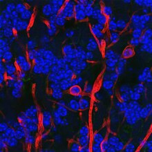
Oligodendrocytes in rat cerebellum stained with antibody to myelin basic protein in red and for DNA in blue. Two oligodendrocyte cell bodies are clearly visible as well as several myelinated axons. These are hollow tubes and so appear as "tramlines" in this confocal image. Most of the DNA is in the nuclei of cerebellum granule cells, which are small interneurons. Image and antibody stain from EnCor Biotechnology Inc.
Myelination is only prevalent in a few brain regions at birth and continues into adulthood. The entire process is not complete until about 25–30 years of age.[20] Myelination is an important component of intelligence, and white matter quantity may be positively correlated with IQ test results in children.[20] Rats that were raised in an enriched environment, which is known to increase cognitive flexibility, had more myelination in their corpus callosi.[21]
It is hypothesized that satellite oligodendrocytes (orperineuronal oligodendrocytes) are functionally distinct from other oligodendrocytes. They are not attached to neurons via myelin sheaths and, therefore, do not contribute to insulation. They remain opposed to neurons and regulate the extracellular fluid.[24] Satellite oligodendrocytes are considered to be a part of the grey matter whereas myelinating oligodendrocytes are a part of the white matter. They may support neuronal metabolism. Satellite oligodendrocytes may be recruited to produce new myelin after a demyelinating injury.[25]
Diseases that result in injury to oligodendrocytes include demyelinating diseases such as multiple sclerosis and various leukodystrophies. Trauma to the body, e.g. spinal cord injury, can also cause demyelination. The immature oligodendrocytes, which increase in number during mid-gestation, are more vulnerable to hypoxic injury and are involved in periventricular leukomalacia.[26] This largely congenital condition of damage to the newly forming brain can therefore lead to cerebral palsy. In cerebral palsy, spinal cord injury, stroke and possibly multiple sclerosis, oligodendrocytes are thought to be damaged by excessive release of the neurotransmitter, glutamate.[27] Damage has also been shown to be mediated by N-methyl-D-aspartate receptors.[27] Oligodendrocyte dysfunction may also be implicated in the pathophysiologyofschizophrenia and bipolar disorder.[28]
Oligodendrocytes are also susceptible to infection by the JC virus, which causes progressive multifocal leukoencephalopathy (PML), a condition that specifically affects white matter, typically in immunocompromised patients. Tumors of oligodendrocytes are called oligodendrogliomas. The chemotherapy agent Fluorouracil (5-FU) causes damage to the oligodendrocytes in mice, leading to both acute central nervous system (CNS) damage and progressively worsening delayed degeneration of the CNS.[29][30]
DNA methylation may also have a role in the degeneration of oligodendrocytes.[31]
^ abCarlson, Neil (2010). Physiology of Behavior. Boston, MA: Allyn & Bacon. pp. 38–39. ISBN978-0-205-66627-0.
^Baumann, Nicole; Pham-Dinh, Danielle (2001-04-01). "Biology of Oligodendrocyte and Myelin in the Mammalian Central Nervous System". Physiological Reviews. 81 (2): 871–927. doi:10.1152/physrev.2001.81.2.871. ISSN0031-9333. PMID11274346.
^Juraska J. M.; Kopcik J. R. (1988). "Sex and environmental influences on the size and ultrastructure of the rat corpus callosum". Brain Research. 450 (1–2): 1–8. doi:10.1016/0006-8993(88)91538-7. PMID3401704. S2CID2720782.
Raine, C.S. (1991). Oligodendrocytes and central nervous system myelin. In Textbook of Neuropathology, second edition, R.L. Davis and D.M. Robertson, eds. (Baltimore, Maryland: Williams and Wilkins), pp. 115–141.