Jump to content


Main menu


Navigation  


Main page
Contents
Current events
Random article
About Wikipedia
Contact us
Donate


Contribute  


Help
Learn to edit
Community portal
Recent changes
Upload file


Search  


Create account

Log in


Create account
 Log in


Pages for logged out editors learn more  


Contributions
Talk


Contents


(Top)


1 Structure  


2 Function  


3 Additional images  


4 References  


5 External links  


Tunica intima


العربية
Български
Deutsch
Español
فارسی
Français
Bahasa Indonesia
Italiano
עברית
Қазақша
Nederlands
Português
Русский
Српски / srpski
Svenska
Українська
 

Edit links


Article
Talk


Read
Edit
View history


Tools


Actions  


Read
Edit
View history


General  


What links here
Related changes
Upload file
Special pages
Permanent link
Page information
Cite this page
Get shortened URL
Download QR code
Wikidata item


Print/export  


Download as PDF
Printable version


In other projects  


Wikimedia Commons


Appearance


From Wikipedia, the free encyclopedia


Tunica intima
Transverse section through a small artery and vein of the mucous membrane of the epiglottis of a child. (Tunica intima is at "e".)
Details
Part ofWall of blood vessels
Identifiers
Latintunica intima
MeSHD017539
TA98A12.0.00.018
TA23922
THH3.09.02.0.01003
FMA55589
Anatomical terminology

[edit on Wikidata]

The tunica intima (Neo-Latin "inner coat"), or intima for short, is the innermost tunica (layer) of an arteryorvein. It is made up of one layer of endothelial cells and is supported by an internal elastic lamina. The endothelial cells are in direct contact with the blood flow.

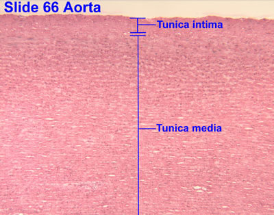
The three layers of a blood vessel are an inner layer (the tunica intima), a middle layer (the tunica media), and an outer layer (the tunica externa).

Indissection, the inner coat (tunica intima) can be separated from the middle (tunica media) by a little maceration, or it may be stripped off in small pieces; but, because of its friability, it cannot be separated as a complete membrane. It is a fine, transparent, colorless structure which is highly elastic, and, after death, is commonly corrugated into longitudinal wrinkles.

Structure[edit]

The structure of the tunica intima depends on the blood vessel type.[1]

Elastic arteries – A single layer of endothelial and a supporting layer of elastin-rich collagen. The layer also contains fibroblasts and smooth muscle cells called 'myointimal cells'

Muscular arteriesEndothelial cells

Arterioles – A single layer of endothelial cells

VeinsEndothelial cells[2]

The inner coat consists of:

  1. A layer of pavement endothelium, the cells of which are polygonal, oval, or fusiform, and have very distinct round or oval nuclei. This endothelium is brought into view most distinctly by staining with silver nitrate.
  2. A subendothelial layer, consisting of delicate connective tissue with branched cells lying in the interspaces of the tissue; in arteries of less than 2 mm in diameter the subendothelial layer consists of a single stratum of stellate cells, and the connective tissue is only largely developed in vessels of a considerable size.[citation needed]
  3. An elastic or fenestrated layer, which consists of a membrane containing a network of elastic fibers, having principally a longitudinal direction, and in which, under the microscope, small elongated apertures or perforations may be seen, giving it a fenestrated appearance. It was therefore called by Henle the fenestrated membrane. This membrane forms the chief thickness of the inner coat, and can be separated into several layers, some of which present the appearance of a network of longitudinal elastic fibers, and others a more membranous character, marked by pale lines having a longitudinal direction. In minute arteries the fenestrated membrane is a very thin layer; but in the larger arteries, and especially in the aorta, it has a considerable thickness.

Function[edit]

Endothelium had been seen to be simply the boundary between the blood in the lumen and the walls of the vessels. However, endothelium has been shown to release local chemicals called endothelins which are powerful vasoconstrictors.[3] Endothelins help to regulate capillary exchange and alter blood flow by their constriction of the smooth muscle in the walls. Vasoconstriction increases blood pressure, and its overexpression can contribute to hypertension and cardiovascular disease.[4]

Additional images[edit]

References[edit]

Public domain This article incorporates text in the public domain from page 498 of the 20th edition of Gray's Anatomy (1918)

  1. ^ Steve, Paxton; Michelle, Peckham; Adele, Knibbs (2003). "The Leeds Histology Guide". {{cite journal}}: Cite journal requires |journal= (help)
  • ^ Steve, Paxton; Michelle, Peckham; Adele, Knibbs (2003). "The Leeds Histology Guide". {{cite journal}}: Cite journal requires |journal= (help)
  • ^ Haryono, A; Ramadhiani, R; Ryanto, GRT; Emoto, N (16 May 2022). "Endothelin and the Cardiovascular System: The Long Journey and Where We Are Going". Biology. 11 (5): 759. doi:10.3390/biology11050759. PMC 9138590. PMID 35625487.
  • ^  This article incorporates text available under the CC BY 4.0 license. Betts, J Gordon; Desaix, Peter; Johnson, Eddie; Johnson, Jody E; Korol, Oksana; Kruse, Dean; Poe, Brandon; Wise, James; Womble, Mark D; Young, Kelly A (June 8, 2023). Anatomy & Physiology. Houston: OpenStax CNX. 20.1 Structure and function of blood vessles. ISBN 978-1-947172-04-3.
  • External links[edit]


    Retrieved from "https://en.wikipedia.org/w/index.php?title=Tunica_intima&oldid=1222042420"

    Categories: 
    Wikipedia articles incorporating text from the 20th edition of Gray's Anatomy (1918)
    Arteries
    Hidden categories: 
    CS1 errors: missing periodical
    Articles with imported Creative Commons Attribution 4.0 text
    Articles with short description
    Short description matches Wikidata
    Articles lacking in-text citations from May 2015
    All articles lacking in-text citations
    All articles with unsourced statements
    Articles with unsourced statements from May 2017
    Articles with TA98 identifiers


    This page was last edited on 3 May 2024, at 14:43 (UTC).

    Text is available under the Creative Commons Attribution-ShareAlike License 4.0; additional terms may apply. By using this site, you agree to the Terms of Use and Privacy Policy. Wikipedia® is a registered trademark of the Wikimedia Foundation, Inc., a non-profit organization.


    Privacy policy

    About Wikipedia

    Disclaimers

    Contact Wikipedia

    Code of Conduct

    Developers

    Statistics

    Cookie statement

    Mobile view


    Wikimedia Foundation
    Powered by MediaWiki