
チン小帯
| チン小帯 | |
|---|---|
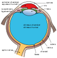
 ヒトの目の模式図(チン小帯は、図の左上に描かれている。) | |
 眼球の矢状面の上半分(チン小帯は、中央付近に見える。) | |
| 概要 | |
| 表記・識別 | |
| ラテン語 | zonula ciliaris |
| ドーランド /エルゼビア | z_01/12870397 |
| グレイ解剖学 | p.1018 |
| TA | A15.2.05.015 |
| FMA | 58838 |
| 解剖学用語 | |
チン小帯︵チンしょうたい、Zinn’s membrane, ciliary zonule︶は、毛様体小帯または毛様小帯とも呼び、毛様体を水晶体に繋ぐ環状の線維である。ヨハン・ゴットフリート・ツィンにちなんで名づけられた。
はたらき[編集]
チン小帯は眼球の内部で水晶体を固定している、線維のあつまりである[1]。チン小帯が水晶体を支えることによって、水晶体はその形を左右対称に保ち、ピントを網膜に合わせることができる[1]。加齢にしたがって劣化していくほか、目をこすることによっても劣化し[1]、一旦切れると再生しない[1]。発達[編集]
毛様体上皮細胞がチン小帯の一部を形成している可能性がある[2]。解剖学[編集]
チン小帯は、2つの層に分かれている。薄い層は硝子体窩に並び、厚い層は小帯線維が集まっている。線維は、毛様体小体として知られる[3]。チン小帯の直径は、約1から2 μmである[4]。 水晶体との摩擦等で、色素顆粒がチン小帯から放出されると、虹彩の色は徐々に薄くなる。これらの色素顆粒がチャネルを阻害し、緑内障を引き起こすこともある。 チン小帯は、主に結合組織タンパク質であるフィブリリンで構成されている[2]。フィブリリン遺伝子の変異はマルファン症候群を引き起こし、水晶体脱臼の危険が高まる[2]。見かけ[編集]
チン小帯は、細隙灯で見ることは難しいが、子供のものや虹彩欠損症、水晶体亜脱臼の場合は見えることがある[5]。チン小帯の数は、年齢とともに減少する[4]。チン小帯は、水晶体外縁の前方及び後方に挿入されているように見える[6]。画像[編集]
-
 目の模式図
目の模式図 -
 目の模式図
目の模式図


出典[編集]
- ^ a b c d 赤星, 隆幸 (2019年3月16日). “元気のココロ”. 日本経済新聞土曜版 (日本経済新聞): p. S7
- ^ a b c Kaufman, Paul L.; Alm, Albert (2010). Adler's physiology of the eye (11th ed. ed.). St. Louis, Mo: Mosby. pp. 145-146. ISBN 978-0-323-05714-1
- ^ http://www.etsu.edu/cpah/hsci/forsman/WebVision.htm
- ^ a b Bornfeld, Norbert; Spitznas, Manfred; Breipohl, Winrich; Bijvank, Gerhard J. (1974). “Scanning electron microscopy of the zonule of Zinn”. Albrecht von Graefes Archiv for Klinische und Experimentelle Ophthalmologie 192 (2): 117-129. doi:10.1007/BF00410698.
- ^ McCulloch, C (1954-1955). “The Zonule of Zinn: its Origin, Course, and Insertion, and its Relation to Neighboring Structures.”. Transactions of the American Ophthalmological Society 52: 525-85. PMID 13274438 2012年12月15日閲覧。.
- ^ Farnsworth, PN; Mauriello, JA; Burke-Gadomski, P; Kulyk, T; Cinotti, AA (1976 Jan). “Surface ultrastructure of the human lens capsule and zonular attachments.”. Investigative ophthalmology 15 (1): 36-40. PMID 1245377.
外部リンク[編集]
- Diagram at unmc.edu
- Diagram at eye-surgery-uk.com
- Diagram and overview at webschoolsolutions.com
- ciliary+zonule - eMedicine Dictionary
- Histology image: 08011loa — ボストン大学の組織学学習システム