Jump to content


Main menu


Navigation  


Main page
Contents
Current events
Random article
About Wikipedia
Contact us
Donate


Contribute  


Help
Learn to edit
Community portal
Recent changes
Upload file


Search  


Create account

Log in


Create account
 Log in


Pages for logged out editors learn more  


Contributions
Talk


Contents


(Top)


1 Structure  


1.1  Development  


2 Function  


3 Clinical significance  


4 History  


5 Other animals  


6 Additional images  


7 See also  


8 References  


Malleus


العربية
Azərbaycanca
 / Bân-lâm-gú
Башҡортса
Bosanski
Brezhoneg
Català
Чӑвашла
Čeština
Corsu
Cymraeg
Dansk
Deutsch
Español
Esperanto
Euskara
فارسی
Français
Galego

Hrvatski
Ido
Bahasa Indonesia
Italiano
Қазақша
Кыргызча
Latina
Македонски

Nederlands
 

Norsk bokmål
Norsk nynorsk
Polski
Português
Română
Русский
Slovenčina
Српски / srpski
Srpskohrvatski / српскохрватски
Suomi
Svenska
ி


Türkçe
Українська


Edit links


Article
Talk


Read
Edit
View history


Tools


Actions  


Read
Edit
View history


General  


What links here
Related changes
Upload file
Special pages
Permanent link
Page information
Cite this page
Get shortened URL
Download QR code
Wikidata item


Print/export  


Download as PDF
Printable version


In other projects  


Wikimedia Commons


Appearance


From Wikipedia, the free encyclopedia


Malleus
Left malleus. A. From behind. B. From within.
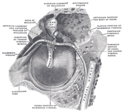
The right membrana tympani with the hammer and the chorda tympani, viewed from within, from behind, and from above (malleus visible at center)
Details
Pronunciation/ˈmæliəs/
PrecursorFirst branchial arch
Part ofMiddle ear
SystemAuditory system
Identifiers
Latinmalleus
MeSHD008307
TA98A15.3.02.043
TA2881
FMA52753
Anatomical terms of bone

[edit on Wikidata]


The malleus, or hammer, is a hammer-shaped small bone or ossicle of the middle ear. It connects with the incus, and is attached to the inner surface of the eardrum. The word is Latin for 'hammer' or 'mallet'. It transmits the sound vibrations from the eardrum to the incus (anvil).

Structure[edit]

The malleus is a bone situated in the middle ear. It is the first of the three ossicles, and attached to the tympanic membrane. The head of the malleus is the large protruding section, which attaches to the incus. The head connects to the neck of malleus. The bone continues as the handle (or manubrium) of malleus, which connects to the tympanic membrane.[1] Between the neck and handle of the malleus, lateral and anterior processes emerge from the bone.[2][3] The bone is oriented so that the head is superior and the handle is inferior.[3]

Development[edit]

Embryologically, the malleus is derived from the first pharyngeal arch along with the incus.[3] It grows from Meckel's cartilage.[3]

Function[edit]

The malleus is one of three ossicles in the middle ear which transmit sound from the tympanic membrane (ear drum) to the inner ear. The malleus receives vibrations from the tympanic membrane and transmits this to the incus.[2]

Clinical significance[edit]

The malleus may be palpatedbysurgeons during ear surgery.[1] It may become fixed in place due to surgical complications, causing hearing loss.[1] This may be corrected with further surgery.[1]

History[edit]

Several sources attribute the discovery of the malleus to the anatomist and philosopher Alessandro Achillini.[4][5] The first brief written description of the malleus was by Berengario da Carpi in his Commentaria super anatomia Mundini (1521).[6] Niccolo Massa's Liber introductorius anatomiae[7] described the malleus in slightly more detail and likened both it and the incus to little hammers terming them malleoli.[8]

Other animals[edit]

The malleus is unique to mammals, and evolved from a lower jaw bone in basal amniotes called the articular, which still forms part of the jaw joint in reptiles and birds.[9][10]

Additional images[edit]

  • Head and neck of a human embryo eighteen weeks old, with Meckel's cartilage and hyoid bone exposed.
    Head and neck of a human embryo eighteen weeks old, with Meckel's cartilage and hyoid bone exposed.
  • External and middle ear, opened from the front. Right side.
    External and middle ear, opened from the front. Right side.
  • Chain of ossicles and their ligaments, seen from the front in a vertical, transverse section of the tympanum.
    Chain of ossicles and their ligaments, seen from the front in a vertical, transverse section of the tympanum.
  • CT image of malleus
    CT image of malleus
  • Auditory ossicles. Tympanic cavity. Deep dissection.
    Auditory ossicles. Tympanic cavity. Deep dissection.
  • Auditory ossicles. Incus and malleus. Deep dissection.
    Auditory ossicles. Incus and malleus. Deep dissection.
  • See also[edit]

    References[edit]

    1. ^ a b c d Lippy, William H.; Berenholz, Leonard P. (2010). "25 - Special Problems of Otosclerosis Surgery". Otologic surgery (3rd ed.). Philadelphia: Saunders. pp. 293–303. doi:10.1016/B978-1-4160-4665-3.00025-1. ISBN 978-1-4377-1966-6. OCLC 489078311.
  • ^ a b Mitchell, Richard L. Drake, Wayne Vogl, Adam W. M. (2005). Gray's anatomy for students. Philadelphia, Pa.: Elsevier. p. 862. ISBN 978-0-8089-2306-0.{{cite book}}: CS1 maint: multiple names: authors list (link)
  • ^ a b c d Cunningham, Craig (2016). "5 - The Skull". Developmental juvenile osteology. Louise Scheuer, Sue M. Black, Helen Liversidge, Angela Christie (2nd ed.). Amsterdam: Academic Press. pp. 43–148. doi:10.1016/B978-0-12-382106-5.00005-0. ISBN 978-0-12-382107-2. OCLC 956277358.
  • ^ Alidosi, GNP. I dottori Bolognesi di teologia, filosofia, medicina e d'arti liberali dall'anno 1000 per tutto marzo del 1623, Tebaldini, N., Bologna, 1623. http://gallica.bnf.fr/ark:/12148/bpt6k51029z/f35.image#
  • ^ Lind, L. R. Studies in pre-Vesalian anatomy. Biography, translations, documents, American Philosophical Society, Philadelphia, 1975. p.40
  • ^ Jacopo Berengario da Carpi,Commentaria super anatomia Mundini, Bologna, 1521. https://archive.org/details/ita-bnc-mag-00001056-001
  • ^ Niccolo Massa, Liber introductorius anatomiae, Venice, 1536. p.166. https://www.digitale-sammlungen.de/en/view/bsb10151904?page=1
  • ^ O'Malley, C.D. Andreas Vesalius of Brussels, 1514-1564. Berkeley: University of California Press, 1964. p. 120
  • ^ Ramachandran, V. S.; Blakeslee, S. (1999). Phantoms in the Brain. Quill. pp. 210. ISBN 9780688172176.
  • ^ Luo, Zhe-Xi (2021). "2.14 - Origins and Early Evolution of Mammalian Ears and Hearing Function". The Senses: A Comprehensive Reference. Vol. 2. Bernd Fritzsch (2nd ed.). Cambridge,Massachusetts: Academic Press. pp. 207–252. doi:10.1016/B978-0-12-805408-6.00033-6. ISBN 978-0-12-805409-3. OCLC 1196340700.

  • Retrieved from "https://en.wikipedia.org/w/index.php?title=Malleus&oldid=1223990491"

    Categories: 
    Auditory system
    Bones of the head and neck
    Ear
    Ossicles
    Otorhinolaryngology
    Otology
    Middle ear
    Hidden categories: 
    CS1 maint: multiple names: authors list
    Articles with short description
    Short description is different from Wikidata
    Articles with J9U identifiers
    Articles with LCCN identifiers
    Articles with TA98 identifiers


    This page was last edited on 15 May 2024, at 16:08 (UTC).

    Text is available under the Creative Commons Attribution-ShareAlike License 4.0; additional terms may apply. By using this site, you agree to the Terms of Use and Privacy Policy. Wikipedia® is a registered trademark of the Wikimedia Foundation, Inc., a non-profit organization.


    Privacy policy

    About Wikipedia

    Disclaimers

    Contact Wikipedia

    Code of Conduct

    Developers

    Statistics

    Cookie statement

    Mobile view


    Wikimedia Foundation
    Powered by MediaWiki