Jump to content


Main menu


Navigation  


Main page
Contents
Current events
Random article
About Wikipedia
Contact us
Donate


Contribute  


Help
Learn to edit
Community portal
Recent changes
Upload file


Search  


Create account

Log in


Create account
 Log in


Pages for logged out editors learn more  


Contributions
Talk


Contents


(Top)


1 Structure  


1.1  Frontal lobe  


1.2  Parietal lobe  


1.3  Temporal lobe  


2 Function  


3 Clinical significance  


3.1  Occlusion  


4 See also  


5 References  


6 External links  


Middle cerebral artery


العربية
Deutsch
Español
فارسی
Français
Galego
Italiano
Latina
Nederlands

Norsk bokmål
Plattdüütsch
Polski
Português
Română
Svenska
Türkçe
Українська
 

Edit links


Article
Talk


Read
Edit
View history


Tools


Actions  


Read
Edit
View history


General  


What links here
Related changes
Upload file
Special pages
Permanent link
Page information
Cite this page
Get shortened URL
Download QR code
Wikidata item


Print/export  


Download as PDF
Printable version


In other projects  


Wikimedia Commons


Appearance


From Wikipedia, the free encyclopedia


Middle cerebral artery
Outer surface of cerebral hemisphere, showing areas supplied by cerebral arteries (Pink is region supplied by middle cerebral artery.)
The arterial circle and arteries of the brain (inferior view). The middle cerebral arteries (top of figure) arise from the internal carotid arteries.
Details
SourceInternal carotid arteries
BranchesAnterolateral central arteries
VeinMiddle cerebral vein
SuppliesCerebrum
Identifiers
Latinarteria cerebri media
MeSHD020768
TA98A12.2.07.046
TA24509
FMA50079
Anatomical terminology

[edit on Wikidata]

The middle cerebral artery (MCA) is one of the three major paired cerebral arteries that supply blood to the cerebrum. The MCA arises from the internal carotid artery and continues into the lateral sulcus where it then branches and projects to many parts of the lateral cerebral cortex. It also supplies blood to the anterior temporal lobes and the insular cortices.

The left and right MCAs rise from trifurcations of the internal carotid arteries and thus are connected to the anterior cerebral arteries and the posterior communicating arteries, which connect to the posterior cerebral arteries. The MCAs are not considered a part of the Circle of Willis.[1]

Structure

[edit]
Middle cerebral artery
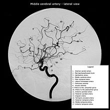
Middle cerebral artery and its branches (patient has a hypoplastic A1 segment and an absent PCOM, resulting in a purely MCA angio from internal carotid artery injection)

The middle cerebral artery divides into four segments, named by the region they supply as opposed to order of branching as the latter can be somewhat variable:[2]

The M2 and M3 segments may each split into 2 or 3 main trunks (terminal branches) with an upper trunk, lower trunk and occasionally a middle trunk. Bifurcations and trifurcations occurs in 50% and 25% of the cases respectively. Other cases include duplication of the MCA at the internal carotid artery (ICA) or an accessory MCA (AccMCA) which arise not from the ICA but as a branch from the anterior cerebral artery.[4] The middle trunk that exist in parts of the population, when present provides the pre-Rolandic, Rolandic, anterior parietal, posterior parietal and the angular artery for irrigation instead of the upper and lower trunks.

The branches of the MCA can be described by the areas that they irrigate.

Frontal lobe

[edit]

Parietal lobe

[edit]

Temporal lobe

[edit]

Function

[edit]

Areas supplied by the middle cerebral artery include:

MCA occlusion site and resulting Aphasia

Clinical significance

[edit]

Occlusion

[edit]

Occlusion of the middle cerebral artery results in Middle cerebral artery syndrome, potentially showing the following defects:

  1. Paralysis (-plegia) or weakness (-paresis) of the contralateral face and arm (faciobrachial)
  2. Sensory loss of the contralateral face and arm.
  3. Damage to the dominant hemisphere (usually the left hemisphere) results in aphasia (i.e. Broca's areaorWernicke's)
  4. Damage to the non-dominant hemisphere (usually the right hemisphere) results in contralateral neglect syndrome, inaccurate localization in the half field, impaired ability to judge distance (nondominant parietal lobe).
  5. Large MCA infarcts often have déviation conjuguée, a gaze preference towards the side of the lesion, especially during the acute period. Contralateral homonymous hemianopsia is often present.

See also

[edit]

References

[edit]
  1. ^ Moore KL, Dalley AR. Clinically Oriented Anatomy, 4th Ed., Lippincott Williams & Wilkins, Toronto. Copyright 1999. ISBN 0-683-06141-0.
  • ^ Krayenbühl, Hugo; Yaşargil, Mahmut Gazi; Huber, Peter; Bosse, George (1982), Cerebral Angiography, Thieme, pp. 105–123, ISBN 978-0-86577-067-6
  • ^ "Middle Cerebral Artery".
  • ^ Osborn, Anne G.; Jacobs, John M. (1999), Diagnostic Cerebral Angiography, Lippincott Williams & Wilkins, pp. 143–144, ISBN 978-0-397-58404-8
  • [edit]
    Retrieved from "https://en.wikipedia.org/w/index.php?title=Middle_cerebral_artery&oldid=1222888049"

    Category: 
    Arteries of the head and neck
    Hidden categories: 
    Articles with short description
    Short description is different from Wikidata
    Anatomy NAV infobox with use of other NAV parameters
    Commons category link from Wikidata
    Articles with TA98 identifiers


    This page was last edited on 8 May 2024, at 15:14 (UTC).

    Text is available under the Creative Commons Attribution-ShareAlike License 4.0; additional terms may apply. By using this site, you agree to the Terms of Use and Privacy Policy. Wikipedia® is a registered trademark of the Wikimedia Foundation, Inc., a non-profit organization.


    Privacy policy

    About Wikipedia

    Disclaimers

    Contact Wikipedia

    Code of Conduct

    Developers

    Statistics

    Cookie statement

    Mobile view


    Wikimedia Foundation
    Powered by MediaWiki