Jump to content


Main menu


Navigation  


Main page
Contents
Current events
Random article
About Wikipedia
Contact us
Donate


Contribute  


Help
Learn to edit
Community portal
Recent changes
Upload file


Search  


Create account

Log in


Create account
 Log in


Pages for logged out editors learn more  


Contributions
Talk


Contents


(Top)


1 Structure  


1.1  Contents  


2 Clinical significance  


3 History  


4 Additional images  


5 References  


6 External links  


Adductor canal


العربية
Deutsch
Français

Italiano

Polski
Português
 

Edit links


Article
Talk


Read
Edit
View history


Tools


Actions  


Read
Edit
View history


General  


What links here
Related changes
Upload file
Special pages
Permanent link
Page information
Cite this page
Get shortened URL
Download QR code
Wikidata item


Print/export  


Download as PDF
Printable version


Appearance


From Wikipedia, the free encyclopedia


Adductor canal
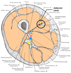
The femoral artery. (Canal not labeled, but region visible at center right.)
Cross-section through the middle of the thigh (the right thigh if seen from below)
Details
Identifiers
Latincanalis adductorius
TA98A04.7.03.006
TA22611
FMA58781
Anatomical terminology

[edit on Wikidata]

The adductor canal (also known as the subsartorial canalorHunter's canal) is an aponeurotic tunnel in the middle third of the thigh giving passage to parts of the femoral artery, vein, and nerve. It extends from the apex of the femoral triangle to the adductor hiatus.

Structure

[edit]

The adductor canal extends from the apex of the femoral triangle to the adductor hiatus. It is an intermuscular cleft situated on the medial aspect of the middle third of the anterior compartment of the thigh, and has the following boundaries:

It is covered by a strong aponeurosis which extends from the vastus medialis, across the femoral vessels to the adductor longus and magnus.

Contents

[edit]

The canal contains the subsartorial artery (distal segment of the femoral artery), subsartorial vein (distal segment of the femoral vein), and branches of the femoral nerve (specifically, the saphenous nerve, and the nerve to the vastus medialis).[1][2][3] The femoral artery with its vein and the saphenous nerve enter this canal through the superior foramen. Then, the saphenous nerve and artery and vein of genus descendens exit through the anterior foramen, piercing the vastoadductor intermuscular septum. Finally, the femoral artery and vein exit via the inferior foramen (usually called the hiatus) through the inferior space between the oblique and medial heads of adductor magnus.[4]

Clinical significance

[edit]

The saphenous nerve may be compressed in the adductor canal.[5] The adductor canal may be accessed for a saphenous nerve block, often used to treat pain caused by this compression.[5]

History

[edit]

The eponym "Hunter's canal" is named for John Hunter.[6][7]

Additional images

[edit]

References

[edit]

Public domain This article incorporates text in the public domain from page 627 of the 20th edition of Gray's Anatomy (1918)

  1. ^ Amarnath C and Hemant Patel (2023). Comprehensive Textbook of Clinical Radiology - Volume III: Chest and Cardiovascular system. Elsevier Health Sciences. ISBN 9788131263617. Page 1072
  • ^ Sauerland, Eberhardt K.; Patrick W. Tank; Tank, Patrick W. (2005). Grant's dissector. Hagerstown, MD: Lippincott Williams & Wilkins. p. 128. ISBN 0-7817-5484-4.
  • ^ Sambrook, Philip (2010-01-01), Sambrook, Philip; Schrieber, Leslie; Taylor, Thomas; Ellis, Andrew M. (eds.), "Bone Structure and Function in Normal and Disease States", The Musculoskeletal System (Second Edition), Churchill Livingstone, pp. 61–76, doi:10.1016/b978-0-7020-3377-3.00005-6, ISBN 978-0-7020-3377-3, retrieved 2021-02-18
  • ^ А.В. Николаев. (2007). Топографическая анатомия и оперативная хирургия. Москва: ГЭОТАР-Медиа (GEOTAR-Media). pp. 174–176. ISBN 978-5-9704-1207-7.
  • ^ a b Zhang, Yi (2011-01-01), Lennard, Ted A.; Walkowski, Stevan; Singla, Aneesh K.; Vivian, David G. (eds.), "27 - Lower Extremity: Saphenous Nerve Block", Pain Procedures in Clinical Practice (Third Edition), Saint Louis: Hanley & Belfus, pp. 267–270, doi:10.1016/b978-1-4160-3779-8.10027-2, ISBN 978-1-4160-3779-8, retrieved 2021-02-18
  • ^ synd/105atWho Named It?
  • ^ "CHAPTER 15: THE THIGH AND KNEE". Archived from the original on 21 January 2008. Retrieved 2008-01-27.
  • [edit]
    Retrieved from "https://en.wikipedia.org/w/index.php?title=Adductor_canal&oldid=1219076300"

    Categories: 
    Wikipedia articles incorporating text from the 20th edition of Gray's Anatomy (1918)
    Anatomy
    Hidden categories: 
    Articles with short description
    Short description is different from Wikidata
    Articles with TA98 identifiers


    This page was last edited on 15 April 2024, at 16:14 (UTC).

    Text is available under the Creative Commons Attribution-ShareAlike License 4.0; additional terms may apply. By using this site, you agree to the Terms of Use and Privacy Policy. Wikipedia® is a registered trademark of the Wikimedia Foundation, Inc., a non-profit organization.


    Privacy policy

    About Wikipedia

    Disclaimers

    Contact Wikipedia

    Code of Conduct

    Developers

    Statistics

    Cookie statement

    Mobile view


    Wikimedia Foundation
    Powered by MediaWiki