Jump to content


Main menu


Navigation  


Main page
Contents
Current events
Random article
About Wikipedia
Contact us
Donate


Contribute  


Help
Learn to edit
Community portal
Recent changes
Upload file


Search  


Create account

Log in


Create account
 Log in


Pages for logged out editors learn more  


Contributions
Talk


Contents


(Top)


1 Structure  


2 Clinical significance  


2.1  Fracture of distal femur  


2.2  Popliteal artery entrapment syndrome  


3 Additional images  


4 References  


Adductor hiatus


Deutsch
Français
 

Edit links


Article
Talk


Read
Edit
View history


Tools


Actions  


Read
Edit
View history


General  


What links here
Related changes
Upload file
Special pages
Permanent link
Page information
Cite this page
Get shortened URL
Download QR code
Wikidata item


Print/export  


Download as PDF
Printable version


Appearance


From Wikipedia, the free encyclopedia


Adductor hiatus
Deep muscles of the medial femoral region. (Adductor hiatus visible as hole in adductor magnus at lower left.)
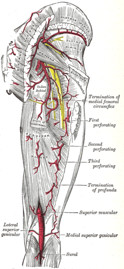
The arteries of the gluteal and posterior femoral regions. (Adductor hiatus is not labeled, but popliteal artery is visible at bottom center.)
Details
Identifiers
Latinhiatus adductorius
TA98A04.7.03.008
TA22634
FMA58784
Anatomical terminology

[edit on Wikidata]

In human anatomy, the adductor hiatus also known as hiatus magnus is a hiatus (gap) between the adductor magnus muscle and the femur that allows the passage of the femoral vessels from the anterior thigh to the posterior thigh and then the popliteal fossa.[1] It is the termination of the adductor canal and lies about 8–13.5 cm (3.1–5.3 in) superior to the adductor tubercle.[2]

Structure

[edit]

Kale et al. classified the adductor hiatus according to its shape and the structures surrounding.[3] An adductor hiatus is described as oval or bridging depending on the shape of the upper boundary. It can also be described as muscularorfibrous depending on whether the structure surrounding is the muscular part or the tendinous part of the adductor magnus muscle. For example, the top drawing on the right shows an oval fibrous type of adductor hiatus, and the bottom one shows a bridging muscular adductor hiatus.

Four structures are associated with the adductor hiatus. However, only two structures enter and then leave through the hiatus; namely the femoral artery and femoral vein. Those vessels become the popliteal vessels (popliteal artery and popliteal vein) immediately after they leave the hiatus,[4] where they form a network of anastomoses called the genicular arteries. The genicular arteries supply the knee joint.

The other two structures that are associated with the adductor hiatus are the descending genicular artery and the saphenous nerve. The saphenous nerve does not leave through the adductor hiatus but penetrates superficially halfway through the adductor canal.

Clinical significance

[edit]

Fracture of distal femur

[edit]

Fracture at the supracondylar area of femur, where the adductor part of the adductor magnus attaches, will most likely cause damage to the femoral artery and may cause impairment of the blood supply to the lower leg.[5] Popliteal artery can also be damaged by the fracture of distal femur.[6]

Popliteal artery entrapment syndrome

[edit]

Abnormality in the relationship between the adductor hiatus and the popliteal artery can also contribute to a condition called popliteal artery entrapment syndrome.[7]

Additional images

[edit]

References

[edit]
  1. ^ Moore, Keith L.; Dalley, Arthur F.; Agur, A. M. R. (2013-02-13). Clinically Oriented Anatomy. Lippincott Williams & Wilkins. ISBN 9781451119459.
  • ^ Olson, S. A.; Holt, B. T. (Feb 1995). "Anatomy of the medial distal femur: a study of the adductor hiatus". Journal of Orthopaedic Trauma. 9 (1): 63–65. doi:10.1097/00005131-199502000-00010. ISSN 0890-5339. PMID 7714656.
  • ^ Kale, Ayşin; Gayretli, Ozcan; Oztürk, Adnan; Gürses, Ilke Ali; Dikici, Fatih; Usta, Ahmet; Sahinoğlu, Kayıhan (Dec 2012). "Classification and localization of the adductor hiatus: a cadaver study". Balkan Medical Journal. 29 (4): 395–400. doi:10.5152/balkanmedj.2012.030. ISSN 2146-3123. PMC 4115878. PMID 25207041.
  • ^ Agur, A. M. R.; Dalley, Arthur F.; Grant, John Charles Boileau (2013). Grant's Atlas of Anatomy. Lippincott Williams & Wilkins. ISBN 9781608317561.
  • ^ Copley, L. A.; Dormans, J. P.; Davidson, R. S. (1996). "Vascular injuries and their sequelae in pediatric supracondylar humeral fractures: toward a goal of prevention". Journal of Pediatric Orthopedics. 16 (1): 99–103. doi:10.1097/00004694-199601000-00020. ISSN 0271-6798. PMID 8747364.
  • ^ Green, Neil E.; Swiontkowski, Marc F. (2009). Skeletal Trauma in Children. Elsevier Health Sciences. ISBN 978-1416049005.
  • ^ Wright, Lonnie B.; Matchett, W. Jean; Cruz, Carlos P.; James, Charles A.; Culp, William C.; Eidt, John F.; McCowan, Timothy C. (March 1, 2004). "Popliteal Artery Disease: Diagnosis and Treatment". RadioGraphics. 24 (2): 467–479. doi:10.1148/rg.242035117. ISSN 0271-5333. PMID 15026594.

  • Retrieved from "https://en.wikipedia.org/w/index.php?title=Adductor_hiatus&oldid=1188213334"

    Category: 
    Human anatomy
    Hidden categories: 
    Articles with short description
    Short description matches Wikidata
    Articles with TA98 identifiers


    This page was last edited on 4 December 2023, at 00:30 (UTC).

    Text is available under the Creative Commons Attribution-ShareAlike License 4.0; additional terms may apply. By using this site, you agree to the Terms of Use and Privacy Policy. Wikipedia® is a registered trademark of the Wikimedia Foundation, Inc., a non-profit organization.


    Privacy policy

    About Wikipedia

    Disclaimers

    Contact Wikipedia

    Code of Conduct

    Developers

    Statistics

    Cookie statement

    Mobile view


    Wikimedia Foundation
    Powered by MediaWiki