Jump to content


Main menu


Navigation  


Main page
Contents
Current events
Random article
About Wikipedia
Contact us
Donate


Contribute  


Help
Learn to edit
Community portal
Recent changes
Upload file


Search  


Create account

Log in


Create account
 Log in


Pages for logged out editors learn more  


Contributions
Talk


Contents


(Top)


1 Additional images  


2 See also  


3 References  


Inferior extensor retinaculum of foot


Français


Edit links


Article
Talk


Read
Edit
View history


Tools


Actions  


Read
Edit
View history


General  


What links here
Related changes
Upload file
Special pages
Permanent link
Page information
Cite this page
Get shortened URL
Download QR code
Wikidata item


Print/export  


Download as PDF
Printable version


Appearance


From Wikipedia, the free encyclopedia


Inferior extensor retinaculum of foot
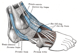
The mucous sheaths of the tendons around the ankle. Lateral aspect.
The mucous sheaths of the tendons around the ankle. Medial aspect.
Details
Identifiers
Latinretinaculum musculorum extensorum inferius pedis, ligamentum cruciatum cruris
TA98A04.7.03.027
TA22713
FMA49385
Anatomical terminology

[edit on Wikidata]

The inferior extensor retinaculum of the foot (cruciate crural ligament, lower part of anterior annular ligament) is a Y-shaped band placed in front of the ankle-joint, the stem of the Y being attached laterally to the upper surface of the calcaneus, in front of the depression for the interosseous talocalcaneal ligament; it is directed medialward as a double layer, one lamina passing in front of, and the other behind, the tendons of the peroneus tertius and extensor digitorum longus.[1][2]

At the medial border of the latter tendon, these two layers join, forming a compartment in which the tendons are enclosed.

From the medial extremity of this sheath, the two limbs of the Y diverge: one is directed upward and medialward, to be attached to the tibial malleolus, passing over the extensor hallucis longus and the vessels and nerves but enclosing the tibialis anterior by a splitting of its fibers.

The other limb extends downward and medialward, to be attached to the border of the plantar aponeurosis, and passes over the tendons of the extensor hallucis longus and tibialis anterior and also the vessels and nerves.

Additional images[edit]

See also[edit]

References[edit]

Public domain This article incorporates text in the public domain from page 488 of the 20th edition of Gray's Anatomy (1918)

  1. ^ Gray, Henry; Standring, Susan; Anhand, Neel, eds. (2021). "Chapter 79 Ankle and foot". Gray's Anatomy: the anatomical basis of clinical practice (42nd ed.). Amsterdam: Elsevier. p. 1430. ISBN 978-0-7020-7705-0.
  • ^ Dalmau-Pastor, M.; Yasui, Y.; Calder, J. D.; Karlsson, J.; Kerkhoffs, G. M. M. J.; Kennedy, J. G. (2016-05-29). "Anatomy of the inferior extensor retinaculum and its role in lateral ankle ligament reconstruction: a pictorial essay". Knee Surgery, Sports Traumatology, Arthroscopy. 24 (4): 957–962. doi:10.1007/s00167-016-4082-5. hdl:2445/110811. ISSN 0942-2056 – via Springer.

  • Retrieved from "https://en.wikipedia.org/w/index.php?title=Inferior_extensor_retinaculum_of_foot&oldid=1224311640"

    Categories: 
    Wikipedia articles incorporating text from the 20th edition of Gray's Anatomy (1918)
    Lower limb anatomy
    Hidden categories: 
    Articles with short description
    Short description matches Wikidata
    Articles with TA98 identifiers


    This page was last edited on 17 May 2024, at 16:12 (UTC).

    Text is available under the Creative Commons Attribution-ShareAlike License 4.0; additional terms may apply. By using this site, you agree to the Terms of Use and Privacy Policy. Wikipedia® is a registered trademark of the Wikimedia Foundation, Inc., a non-profit organization.


    Privacy policy

    About Wikipedia

    Disclaimers

    Contact Wikipedia

    Code of Conduct

    Developers

    Statistics

    Cookie statement

    Mobile view


    Wikimedia Foundation
    Powered by MediaWiki