J u m p t o c o n t e n t
M a i n m e n u
M a i n m e n u
N a v i g a t i o n
● M a i n p a g e ● C o n t e n t s ● C u r r e n t e v e n t s ● R a n d o m a r t i c l e ● A b o u t W i k i p e d i a ● C o n t a c t u s ● D o n a t e
C o n t r i b u t e
● H e l p ● L e a r n t o e d i t ● C o m m u n i t y p o r t a l ● R e c e n t c h a n g e s ● U p l o a d f i l e
S e a r c h
Search
A p p e a r a n c e
● C r e a t e a c c o u n t ● L o g i n
P e r s o n a l t o o l s
● C r e a t e a c c o u n t ● L o g i n
P a g e s f o r l o g g e d o u t e d i t o r s l e a r n m o r e ● C o n t r i b u t i o n s ● T a l k
( T o p )
1 S e e a l s o
2 S o u r c e s a n d n o t e s
T o g g l e t h e t a b l e o f c o n t e n t s
M 1 p r o t e i n
3 l a n g u a g e s
● E s p a ñ o l ● F r a n ç a i s ● 日 本 語
E d i t l i n k s
● A r t i c l e ● T a l k
E n g l i s h
● R e a d ● E d i t ● V i e w h i s t o r y
T o o l s
T o o l s
A c t i o n s
● R e a d ● E d i t ● V i e w h i s t o r y
G e n e r a l
● W h a t l i n k s h e r e ● R e l a t e d c h a n g e s ● U p l o a d f i l e ● S p e c i a l p a g e s ● P e r m a n e n t l i n k ● P a g e i n f o r m a t i o n ● C i t e t h i s p a g e ● G e t s h o r t e n e d U R L ● D o w n l o a d Q R c o d e ● W i k i d a t a i t e m
P r i n t / e x p o r t
● D o w n l o a d a s P D F ● P r i n t a b l e v e r s i o n
A p p e a r a n c e
F r o m W i k i p e d i a , t h e f r e e e n c y c l o p e d i a
The M1 protein binds to the viral RNA . The binding is not specific to any RNA sequence, and is performed via a peptide sequence rich in basic amino acids .[citation needed
It also has multiple regulatory functions, performed by interaction with the components of the host cell. The mechanisms regulated include a role in the export of the viral ribonucleoproteins from the host cell nucleus , inhibition of viral transcription , and a role in the virus assembly and budding . The protein was found to undergo phosphorylation in the host cell.[citation needed
The M1 protein forms a layer under the patches of host cell membrane that are rich with the viral hemagglutinin , neuraminidase and M2 transmembrane proteins , and facilitates budding of the mature viruses.[citation needed
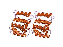
M1 consists of two domains connected by a linker sequence . The N-terminal domain has a multi-helical structure that can be divided into two subdomains.[2] alpha-helical structure .
See also
[ edit ]
Sources and notes
[ edit ]
^ Arzt S, Baudin F, Barge A, Timmins P, Burmeister WP, Ruigrok RW (January 2001). "Combined results from solution studies on intact influenza virus M1 protein and from a new crystal form of its N-terminal domain show that M1 is an elongated monomer" . Virology . 279 (2 ): 439–46. doi :10.1006/viro.2000.0727 PMID 11162800 .
R e t r i e v e d f r o m " https://en.wikipedia.org/w/index.php?title=M1_protein&oldid=1180942130 " C a t e g o r i e s : ● M e m b r a n e b i o l o g y ● P e r i p h e r a l m e m b r a n e p r o t e i n s ● I n f l u e n z a A v i r u s ● V i r a l s t r u c t u r a l p r o t e i n s H i d d e n c a t e g o r i e s : ● A l l a r t i c l e s w i t h u n s o u r c e d s t a t e m e n t s ● A r t i c l e s w i t h u n s o u r c e d s t a t e m e n t s f r o m A u g u s t 2 0 2 2
● T h i s p a g e w a s l a s t e d i t e d o n 1 9 O c t o b e r 2 0 2 3 , a t 2 0 : 2 3 ( U T C ) . ● T e x t i s a v a i l a b l e u n d e r t h e C r e a t i v e C o m m o n s A t t r i b u t i o n - S h a r e A l i k e L i c e n s e 4 . 0 ;
a d d i t i o n a l t e r m s m a y a p p l y . B y u s i n g t h i s s i t e , y o u a g r e e t o t h e T e r m s o f U s e a n d P r i v a c y P o l i c y . W i k i p e d i a ® i s a r e g i s t e r e d t r a d e m a r k o f t h e W i k i m e d i a F o u n d a t i o n , I n c . , a n o n - p r o f i t o r g a n i z a t i o n . ● P r i v a c y p o l i c y ● A b o u t W i k i p e d i a ● D i s c l a i m e r s ● C o n t a c t W i k i p e d i a ● C o d e o f C o n d u c t ● D e v e l o p e r s ● S t a t i s t i c s ● C o o k i e s t a t e m e n t ● M o b i l e v i e w


