
放射線医学
放射線医学︵ほうしゃせんいがく︶とは、放射線を用いた診断や治療等を中心とした医学の一分野である。
医療機関における診療科名は﹁放射線科﹂とするところが多いが、﹁放射線診断科﹂や﹁放射線治療科﹂を標榜することも可能である。
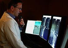
高輝度高精細モニタで画像診断を行う放射線診断医。マイクに口述して いる。

シャウカステンにかけたフィルムで画像診断を行う放射線診断医
Dr. Macintyre's X-Ray Film (1896)


歴史[編集]
この節の加筆が望まれています。 |
1895年、ヴィルヘルム・レントゲン︵Wilhelm Röntgen: 1845.3.27-1923.2.10︶博士はX線を発見し、医療への貢献のみならず近代物理学の幕を開いたこの発見により1901年最初のノーベル物理学賞を受賞した。
X線発見と同じ1895年、早速X線による治療が行われている。文献的に最初に報告されたがんのX線治療は、1896年2月のVoigtによる進行期上咽頭がんの疼痛緩和照射である。
その後、現代では照射装置や治療計画装置の技術的進歩により、強度変調放射線治療 (Intensity-modulated Radiation Therapy: IMRT) や画像誘導放射線治療 (Image-Guided Radiation Therapy) などより高精度な治療へと発展していった。
また、放射線診断学を支える撮影技術に関しても、発展を続け、単純X線写真をはじめとして、1970年代初頭に実用化したラドン変換を基本原理とするコンピュータ断層撮影、1946年のブロッホ、パーセルによる核磁気共鳴信号検出成功に端を発し、1983年に実用化した核磁気共鳴画像法、そして単一光子放出型コンピュータ断層撮影法 (single photon emission computed tomography: SPECT) や陽電子放射断層撮影 (positron emission tomograpy: PET) といったモダリティーが開発されていった。
分類[編集]
大きく以下の三つに分類される。放射線診断学[編集]
●放射線診断学 ●単純X線撮影 ●CT ●MRI ●SPECT ●PET ●超音波検査 ●画像下治療 (Interventional Radiology, IVR) 放射線診断手技を用いた治療法である。直訳すると﹁介入的放射線医学﹂となるが、一般的でなく、﹁IVR﹂、﹁インターベンショナルラジオロジー﹂と呼称されることが通例である[1]が、それが普及の障害になっているとの考えから関連学会で﹁画像下治療﹂と言う訳語が定められた[2]。主に経皮的アプローチにより行なわれ、注射針や細いカテーテルと呼ばれる細い管を用いて血管内から病変部へアプローチするものや、肺や肝臓などに体外から直接針を刺入した上で、病変部の生検をしたりや針先からラジオ波を流すことによるジュール熱によって病変を焼き切ったりするものなど、診断治療への介入は多岐にわたる。従来の手術治療と比べ体への侵襲が少ない方法であり、一部の手術療法の置換による低侵襲化および耐術能に乏しい患者の代替治療の提供を目的として発展してきた。以前は、血管造影や超音波、透視下による治療部位の把握が中心であったが、最近ではCT、MRI等も応用されている[3]。治療対象および方法はかなり広範であり、日進月歩の著しい分野である。治療を行なう部門ではあるが、診断部門に分類される。 ●Vascular IVR ●Non-vascular IVR この他に放射線透視下において、消化器内科的には血管以外に経皮的に胆管を造影したり︵ERCP︶内容物のドレナージを行う︵PTCD︶などの手技や、整形外科的には非観血的に骨や関節を整復するなどの手技がある。放射線治療学[編集]
●放射線療法︵radiation therapy, radiotherapy: RT︶ 高エネルギーのX線、電子線︵electron︶、陽子︵proton︶、重粒子線︵heavy particle︶、中性子︵neutron︶などを照射し、体を突き抜けて悪性細胞に到達し、死滅させるという治療である。 現時点では、主に高エネルギーX線、電子線が用いられており、X線は身体内部の腫瘍、電子線は皮膚がんなどの体表面やそれに近い腫瘍の治療に用いられている。一方で、陽子線治療・重粒子線治療の施設も増えつつあり、また研究レベルでは照射した中性子が薬剤と反応してアルファ線とイオンを放射しがん細胞を傷害するホウ素中性子補足療法︵BNCT︶の施設も設置され始めている。陽子線・重粒子線治療・BNCTの卓越した線量分布は強度変調放射線治療よりも理想に近く、有害事象の軽減が期待されている治療である。 陽子線・重粒子線治療は日本での施設設置当時から2016年ごろまでは、治療技術の確立や治療成績に関する信頼できる論文が少ない事が問題であったが、2022年現在では治療成績に関する論文なども多く報告されている。陽子線治療は2022年現在、小児がんの限局性固形腫瘍、限局性及び局所進行性前立腺がん、頭頸部悪性腫瘍、手術困難な骨軟部腫瘍、4センチメートル以上の切除不能肝細胞がん、切除不能肝内胆管がん、切除不能局所進行膵がん、切除不能局所大腸がん術後再発病変に対して保険収載されている。重粒子線治療では、陽子線治療の保険適用に加えて子宮頸部腺がんが保険適用となっている。核医学[編集]
詳細は「en:Nuclear medicine」および「RI内用療法」を参照
核医学とは、放射性同位元素 (radioisotope; RI) やその化合物の生体内(in vivo)や試験管内(in vitro)の挙動を追跡し、診断・治療を行う医学分野である[4]。核医学画像は、CTやMRIといった他の診断用画像と根本的に異なる側面を持っている。その違いは、CTやMRIは形態画像と呼ばれ、患者の解剖学的な構造を画像に反映するのに対し、核医学画像は機能画像と呼ばれ、種々の放射性薬剤を用いた生理・生化学的機能情報を画像に反映する点にある[5]。
●核医学検査
核医学検査においては、放射性を放出するアイソトープを含んだ薬品(放射性医薬品)を投与し、ガンマカメラ︵シンチカメラまたはアンガー型カメラとも呼ぶ︶で体内での動態を計測する[6]。アイソトープ検査、RI検査ともいわれる。
●核医学における検査・計測条件と目的による分類[7]
●in vivo︵インビボ︶
非密封RIを体内に注射し、各種臓器の機能や動態を直接計測する。
骨シンチグラフィー︵骨シンチ︶や18F-フルオロデオキシグルコース・陽電子放射断層撮影︵18F-FDG-PET:positron emission tomography︶などが、これにあたる。
●in vitro︵インビトロ︶
生体から採取した血液や尿などからホルモンなどの微量物質を生体外で測定する。
核医学による治療
●131Iによって、甲状腺機能亢進症や甲状腺がんのうち、乳頭がんと濾胞がんの治療を行なう。ヨード内用療法。
●90Yによって、一部のリンパ腫の治療を行なう。(商品名‥ゼヴァリン)
●89Srによって、骨シンチで取り込みのある全身性の有痛性多発骨転移に対して、疼痛緩和を図る。︵商品名‥メタストロン︶
●223Raによって、骨転移のある去勢抵抗性前立腺癌に対して、全生存期間︵中央値︶を11.1ヶ月から14ヶ月へ延長する[8]。2016年6月販売開始。︵製品名‥ゾーフィゴ︶
医療被曝[編集]
「被曝」も参照
| 医用画像における実効線量 | |||
|---|---|---|---|
| 対象臓器 | 検査 | 実効線量(大人)[9] | 環境放射線の 等価時間[9] |
| 頭部CT | 単純CT | 2 mSv | 8カ月 |
| 造影剤を使用 | 4 mSv | 16カ月 | |
| 胸部 | 胸部CT | 7 mSv | 2年 |
| 肺がん検診のための胸部CT | 1.5 mSv | 6カ月 | |
| 胸部単純X線撮影 | 0.1 mSv | 10日 | |
| 心臓 | 冠状動脈CT血管造影 | 12 mSv | 4年 |
| 冠状動脈CT、カルシウム走査 | 3 mSv | 1年 | |
| 腹部 | 腹部・骨盤CT | 10 mSv | 3年 |
| 腹部・骨盤CT、低線量プロトコル | 3 mSv[10] | 1年 | |
| 腹部・骨盤CT、造影剤あり | 20 mSv | 7年 | |
| CT結腸検査 | 6 mSv | 2年 | |
| 静脈内腎盂造影 | 3 mSv | 1年 | |
| 上部消化管造影 | 6 mSv | 2年 | |
| 下部消化管造影 | 8 mSv | 3年 | |
| 脊椎 | 脊椎単純X線撮影 | 1.5 mSv | 6カ月 |
| 脊椎CT | 6 mSv | 2年 | |
| 四肢 | 四肢単純X線撮影 | 0.001 mSv | 3時間 |
| 下肢CT血管造影 | 0.3 - 1.6 mSv[11] | 5週間 - 6カ月 | |
| 歯科X線撮影 | 0.005 mSv | 1日 | |
| 骨密度測定(DEXA法) | 0.001 mSv | 3時間 | |
| PET-CT | 25 mSv | 8年 | |
| マンモグラフィー | 0.4 mSv | 7週間 | |

厳密な話をすると、被曝とは、単に身体が電離放射線にさらされたという現象を指す術語である[12]。従って、それに引き続く、何らかの生物学的影響があったとしても、これは被曝とは別個の概念として認識する必要がある。そのため、中立的な概念である被曝により、白内障、唾液分泌低下、粘膜炎、二次発がんなどの患者にとって不利益と考えられる影響や、甲状腺眼症の治療やがんの治癒などの有益と考えられる影響といった相反する事象が併存しても整合性が保たれる。さらに、同じ被曝という事実とそれに引き続く生物学的影響が同じでも、利益・不利益が相対的な場合もある。例えば、被曝すると創傷治癒の遅延や治癒能の低下が生じる。これは一見不利益な影響にも思えるが、これを応用して難治性のケロイドの治療が可能となっている[13]。また全身におおよそ4-10Gy被曝すると骨髄機能が荒廃して致死的となるとされており、この被曝は、患者の著しい不利益につながるとも考えられそうだが、白血病などの血液疾患では、これを応用して骨髄移植前に白血病細胞を死滅させるための前処置として採用されている[14]。
上記のとおり、本来は、言葉の概念上の問題から、生物学的影響の善悪を論じた後、その影響を及ぼす被曝量にさかのぼった上で、被曝の是非を議論されるべきだが、放射線治療分野を除いた、画像診断領域における被曝の生物学的影響は実質的に身体への侵襲と見なして差し支えないことから、通例に従って、以下では両者を特に区別しない。[15]。
医療被曝の線量限度[編集]
放射線防護のため、1990年の国際放射線防護委員会(International Comission on Radiation Protection: ICRP)の勧告に準拠し、日本でも被曝線量限度が法令により定められているが、医療の目的で電離放射線を患者に曝射する場合に限っては、線量限度の法的な定めはない。これは、被曝する本人がその被曝する行為によって診断や治療といった直接の利益を受けるからである[1]。言い換えると、患者が医学的利益を享受する場合には、被曝線量にかかわらず医療被曝が正当化されるということである。これは医療被曝の線量限度を法令で定めてしまうと、国民が適切な医療を受ける機会を失うことと同値である。こうした特殊性から、その他の被曝︵職業被曝・公衆被曝︶と同列に比較されるべきではない。[15]。︵放射線を人体に対して照射する判断は医師および歯科医師のみ可能であり、診療放射線技師は医師又は歯科医師の指示がなければ放射線を人体に対して照射することが許されない。︶ しかしながら結果論ではあるが、放射線診断で健康と診断された場合は被曝という害と健康であるという安心のみが残される事になる。二次予防を目的とした検診における放射線診断では、被曝によるリスクを考慮したガイドラインが設定されている。 ただし、医療被曝の﹁正当化﹂および﹁最適化﹂がなされた上で、被曝が必要最小限となるように行われる必要がある[15]。 放射線医療による、病気の診断・治療を﹁主作用﹂としたとき、医療被曝による生物学的影響のうち好ましくないものを、医療用薬剤になぞらえて﹁副作用﹂とも見なしうる。放射線医療は治療によって患者が得る利益と害(リスク)を考慮して、医師・歯科医師が有益と判断して施される。︵例えば、95%以上の確率で治療が奏功し、回復困難な有害事象を生じる確率が5%以下である、など。︶医療被曝の現状[編集]
放射線診断、放射線治療の進歩と普及に伴い日本を含む一部の医療先進国では医療被曝の実効線量が自然放射線からの被曝より大きくなっている[16]。原子放射線の影響に関する国連科学委員会(UNSCEAR)の2008年の報告によると、全世界での放射線診断は1988年には13.8億回、一人あたりの平均線量は0.35mSvであったが、2008年には31億回、平均線量は0.62mSvとなった。医療先進国の平均は1.92mSvとなっているが日本では2.3mSv[17]、米国は3.0mSv[18]と推定されている。放射線治療に関しては1991~1996年の間は年間470万回であったが、1997~2007年の間では510万回に増加している。直線加速器による治療も増えてきている。医療先進国では放射線治療は1千人あたり年間2.4回(世界平均は0.8回)となっており、頻度は増え続けている。 日本においてはCT機器の普及率が他国より突出しており[19]、人口百万人あたり92.6台(2002年)、2位がオーストラリアで45.3台(2004年)、3位アメリカ32.3台(2004年)であった。この普及率の高さにより、容易に悪く言えば安易に検査を受けることが可能である。CTを1回受けるだけで6.9mSv、胃のX線検査では0.6–2.7mSvの医療被曝がある[20]。 放射線診療における代表的なX線検査での被曝量は、胸部 0.04mSv、腹部1.2mSv、上部消化管 8.7mSv、胸部CT 7.8mSv、腹部CT 7.6mSvである[21][22]。なお、骨髄移植のために行われる全身照射の一回の照射量は2,000mSv(2Gy)で、1日2回の照射を3日間行い、総量で12,000mSv(12Gy)を照射する[23][24]。肺がんに対する定位放射線治療では1回10,000mSv(10Gy)以上の大線量を4回から5回照射して1週間程度で終了させているプロトコルが主流である[25]。不必要な画像診断[編集]
「無駄な医療」も参照
CTのような高額な装置の場合、検査が過剰に行われる懸念が指摘されている[26]。ただ、現時点では、諸外国と比較したときCTとMRIの装置台数は際だって多いが、装置の活用度はほとんど最下位であり、検査数としてはそれほど多いという状況でではない[27]。
実際、低線量の放射線被ばくによる影響には不明な点が多いが、低線量の被ばくも発がんを生じるという仮説︵LNT仮説︶にも基づき15ヵ国で放射線検査の頻度にともなう発癌リスクを調べた結果によれば、日本の医療被曝による発癌リスクは3.2%︵年7587件の発癌数に相当︶と最も高く[28]、これは欧米諸国に比べても3倍程高い数字であり、特徴としてCT検査による被曝が大きな比重を占めており、他国に比べてCT装置の設置台数が多い事などが背景にあるのではと指摘されている[29]。
アメリカ食品医薬品局では画像診断法における不要な放射線照射を減ずる方針が提示されている[30]。一方、被曝を抑えるために装置の改良も行われており、低線量ヘリカルCTなどが開発され、検診対象者をヘビースモーカーといった高リスク群に絞って成果を挙げているが、偽陽性などの問題も指摘されている[31]。
自然被曝[編集]
一方、自然放射線による被曝量は、概ね年間1.0~13mSvの間で世界平均は約2.4mSvである。UNSCEARでは10mSv以上の被曝のある地域を特筆している。 イラン、ブラジル、インドでは、30mSvを超えるようなホットスポットもあり[21][22][32]、インドのケーララ州で家系内遺伝調査をしたところ、高線量地域では統計的に有意に生殖細胞由来の点突然変異が高い傾向にあることが報告されている[33][34]。ブラジルのガラパリでは内部被曝によるものと思われる末梢血リンパ球の染色体異常[35]や、対照地域に比べて癌の死亡率の高さが報告されている[36]が、癌死亡率の報告については他の因子を考慮しておらず、予備的研究の結論とみなすべきだと研究者自身が記述している。しきい線量と影響の事例[編集]
しきい線量 (threshold dose) とは、放射線をある一定レベル以上の被曝を受けると、確定的放射線影響が起きるしきい値となる線量のことであるが[37]、しきい線量のある確定的影響と、しきい線量はないと仮定されている確率的影響とがある[21]。 確定的影響の例には、胎児への影響、器官形成期の被曝による奇形の発生があり、そのしきい線量は100mGy︵ミリグレイ︶とされている[21]。しかしながら、放射線診断での胎児の平均被曝量は、腹部撮影 1.4mGy、注腸造影検査 6.8mGy、腹部CT 8.0mGy、骨盤CT 25.0mGyなどとなっており、このしきい線量100mGyより小さい被曝であり、顕著な影響があるとは考えられないとされている。 確率的影響の例には、複数回のX検査による被曝で白血病またはがんになる可能性がある。米国では、CTスキャンによる検査が年間7000万件以上行われており、そのうちの2万9000件が将来的にCTに関連した癌の発症を引き起こすと推定されている[38]。なお、この確率的影響にはしきい線量はなく、被曝量に比例するとされる。この仮説によると、影響の確率は0にはならないが、日常的な通常の放射線検査での被曝量は、問題となるようなものではないという主張がある。一方、妊娠女性が放射線診断を受ける場合、X線検査の回数と胎児の相対リスクには比例関係があるという報告などもあり[39]、胎児へのリスクをまったく考慮する必要がないとまでは言い切れない不確かさがあり、確定的な結論は出ていない。また、白血病では50〜200mGy以下の被曝では発生率の増加は統計的に明かではない[21]。通常のX線検査では、胸部0.04mGy、腹部0.4mGy、腰椎1.4mGy、上部消化管8.2mGy程度であり、極端な回数の検査をしないかぎり、心配する必要はないという主張もある[21]。脚注[編集]
(一)^ ab南山堂医学大辞典
(二)^ 理事会における IVR の和名についての検討経過概要 (PDF) -日本インターベンショナルラジオロジー学会
(三)^ 新臨床腫瘍学 p.202
(四)^ PETの部屋 核医学の基礎 核医学(Nuclear Medicine)とは - ウェイバックマシン︵2011年9月11日アーカイブ分︶
(五)^ 金原出版 核医学ノート・第4版
(六)^ 核医学会による一般向け説明 (PDF)
(七)^ PETの部屋 核医学の基礎 3つの核医学の分類 - ウェイバックマシン︵2011年9月12日アーカイブ分︶
(八)^ Petrenciuc O‥バイエル薬品社内資料﹇症候性去勢抵抗性前立腺癌患者を対象とした国外第Ⅲ相臨床試験﹈︵2015︶
(九)^ abUnless otherwise specified in boxes, reference is:
- “Radiation Dose in X-Ray and CT Exams”. RadiologyInfo.orgbyRadiological Society of North America. 2017年10月23日閲覧。
(十)^ Brisbane, Wayne; Bailey, Michael R.; Sorensen, Mathew D. (2016). “An overview of kidney stone imaging techniques”. Nature Reviews Urology (Springer Nature) 13 (11): 654–662. doi:10.1038/nrurol.2016.154. ISSN 1759-4812. PMC 5443345.
(11)^ Zhang, Zhuoli; Qi, Li; Meinel, Felix G.; Zhou, Chang Sheng; Zhao, Yan E.; Schoepf, U. Joseph; Zhang, Long Jiang; Lu, Guang Ming (2014). “Image Quality and Radiation Dose of Lower Extremity CT Angiography Using 70 kVp, High Pitch Acquisition and Sinogram-Affirmed Iterative Reconstruction”. PLoS ONE 9 (6): e99112. doi:10.1371/journal.pone.0099112. ISSN 1932-6203.
(12)^ 岩波書店 広辞苑・第六版
(13)^ 金原出版株式会社 良性疾患の放射線治療 p.122
(14)^ 新臨床腫瘍学 p.212
(15)^ abc画像診断ガイドライン 2013年版 p.40
(16)^ 放射線医学総合研究所﹁UNSCEAR2008年報告書﹂ (PDF) 閲覧2011-10-22
(17)^ 文部科学省 ﹁身の回りの放射線﹂ 閲覧2011-10-22
(18)^ Princeton.edu ﹁Background Radiation﹂ - ウェイバックマシン︵2011年10月23日アーカイブ分︶ 閲覧2011-10-22
(19)^ Health at a Glance 2013 (Report). OECD. 21 November 2013. p. 87. doi:10.1787/health_glance-2013-en。
(20)^ 東嶋和子著 ﹃放射線利用の基礎知識﹄ 講談社、2006年12月20日。ISBN 4-06-257518-3。
(21)^ abcdef医療被曝について│聖マリアンナ医科大学
(22)^ ab草間朋子﹃あなたと患者のための放射線防護Q&A﹄医療科学社、ISBN 978-4900770522。
(23)^ この照射方法が主に利用されている(井上俊彦﹁診療 1990年以前の国内における初期の全身照射﹂﹃臨床放射線﹄2008, p.1254)
(24)^ がんの放射線治療──その2全身照射
(25)^ 西村恭昌 ﹃肺がん﹄ 2011, p.90
(26)^ キャサリン・コーフィールド 著、友清裕昭 訳﹃被曝の世紀 放射線の時代に起こったこと﹄朝日新聞社、1990年11月、328-329頁。ISBN 4022562277。"CAT装置は非常に高価である。この支払いのために医師や病院はCATを利用しすぎる傾向が、CATの普及とともに出てきたと心配されている。"。
(27)^ 画像診断ガイドライン 2013年版
(28)^ Amy Berrington de González and Sarah Darby (2004). “Risk of cancer from diagnostic X-rays: estimates for the UK and 14 other countries”. The Lancet 363 (9406): 345-351. doi:10.1016/S0140-6736(04)15433-0.
(29)^ 澤田聡, 渡邉直行, 五十嵐均 (2011). “"Risk of Cancer from Diagnostic X-rays : estimates for the UK and 14 other countries" : Lancet論文レビューと診療放射線技師による放射線防護の立場からのCT検査妥当性についての考察”. 群馬県立県民健康科学大学紀要 6: 73-76. NAID 110008148682. "日本においてCT検査数が多い理由として、CT装置の設置台数が他国に比べてとびぬけて多いという事実がある7)。…︵中略︶…医療機関側に立てば、高額医療機器の導入コストをどう減価償却するかという背景を含んでいる。"
(30)^ FDAが、画像検査による不要な放射線被曝の低減に向けた指針を発表, “海外癌医療情報リファレンス”, 一般社団法人 日本癌医療翻訳アソシエイツ, (2010年2月15日) 2017年10月8日閲覧。
(31)^ 2010/11/16号◆クローズアップ﹁肺癌検診における低線量CTがヘビースモーカーの死亡率に明らかな有効性をもたらす﹂, “海外癌医療情報リファレンス”, 一般社団法人 日本癌医療翻訳アソシエイツ, (2010年11月23日) 2017年10月8日閲覧, "肺癌検診のリスクが潜在的利益と並んで存在し、それも将来の推奨に織り込む必要がある。この試験のスキャン全体の約25%が偽陽性結果を示しており、これは、認められた異常が経過観察時に癌でないと判明したということである。偽陽性と判定された患者は全員、高線量の放射線を用いる診断用CTから肺生検にわたる、何らかの診断法を経過観察時に追加で受け、中には開胸術︵胸部の外科的切開︶を受けた患者もいる。これらはいずれもリスクをもたらすものであると Giaccone氏は説明した。"
(32)^ 世界の高自然放射線地域
(33)^ Lucy Forster et al. (2002). “Natural radioactivity and human mitochondrial DNA mutations”. PNAS 99 (21): 13950-13954. doi:10.1073/pnas.202400499.
(34)^ 翻訳‥伊澤 (2011年5月3日). “自然放射線とヒトミトコンドリア遺伝子の突然変異”. 名古屋生活クラブ. 2011年5月28日閲覧。
(35)^ ブラジルの高自然放射線地域における住民の健康調査 (09-02-07-03) - ATOMICA -
(36)^ Lene H.S. Veigaa and Sérgio Koifman (2005). “Pattern of cancer mortality in some Brazilian HBRAs”. International Congress Series 1276: 110-113. doi:10.1016/j.ics.2004.11.046.
(37)^ しきい線量(threshold dose) - 緊急被ばく医療研修 - ウェイバックマシン︵2013年2月6日アーカイブ分︶
(38)^ Amy Berrington de González et al. (2009). “Projected Cancer Risks From Computed Tomographic Scans Performed in the United States in 2007”. Archives of Internal Medicine 169 (22): 2071-2077. doi:10.1001/archinternmed.2009.440.
(39)^ R. Doll and R. Wakeford (1997). “Risk of childhood cancer from fetal irradiation”. The British Journal of Radiology 70 (830): 130-139. "It is concluded that radiation doses of the order of 10 mGy received by the fetus in utero produce a consequent increase in the risk of childhood cancer."
参考文献[編集]
●﹃南山堂医学大辞典﹄︵第19版︶南山堂、2006年。ISBN 978-4525010294。 ●日本臨床腫瘍学会︵編︶ 編﹃新臨床腫瘍学 がん薬物療法専門医のために﹄︵第4版︶南江堂、2015年。ISBN 978-4-524-26187-1。 ●日本医学放射線学会︵編︶、日本放射線科専門医会・医会︵編︶ 編﹃画像診断ガイドライン 2013年版﹄金原出版、2013年。ISBN 978-4307070935。関連項目[編集]
| 単位 |
| ||||||||
|---|---|---|---|---|---|---|---|---|---|
| 測定 |
| ||||||||
| 放射線の種類 |
| ||||||||
| 物質との相互作用 |
| ||||||||
| 放射線と健康 |
| ||||||||
| 法律・資格 |
| ||||||||
| 関連 |
| ||||||||
| 国立図書館 |
|
|---|---|
| その他 |
|